. 2016 Jul 5;7(27):41053-41066.
doi: 10.18632/oncotarget.9419.

Sialic acid removal from dendritic cells improves antigen cross-presentation and boosts anti-tumor immune responses

Affiliations

Sialic acid removal from dendritic cells improves antigen cross-presentation and boosts anti-tumor immune responses

Mariana Silva et al. Oncotarget. .

Abstract

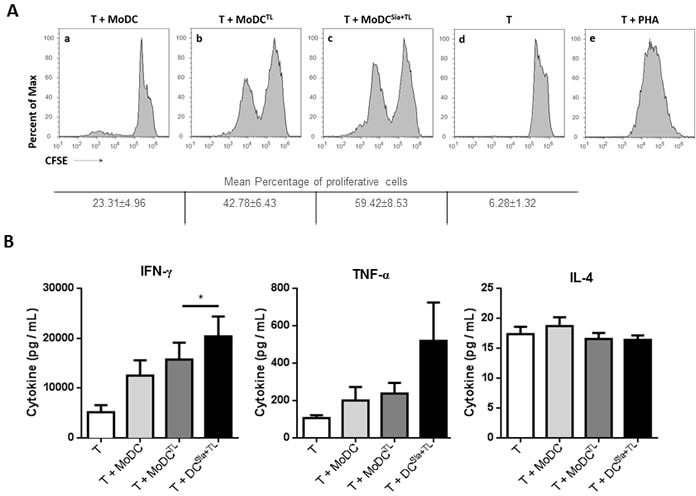
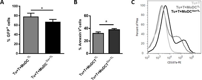
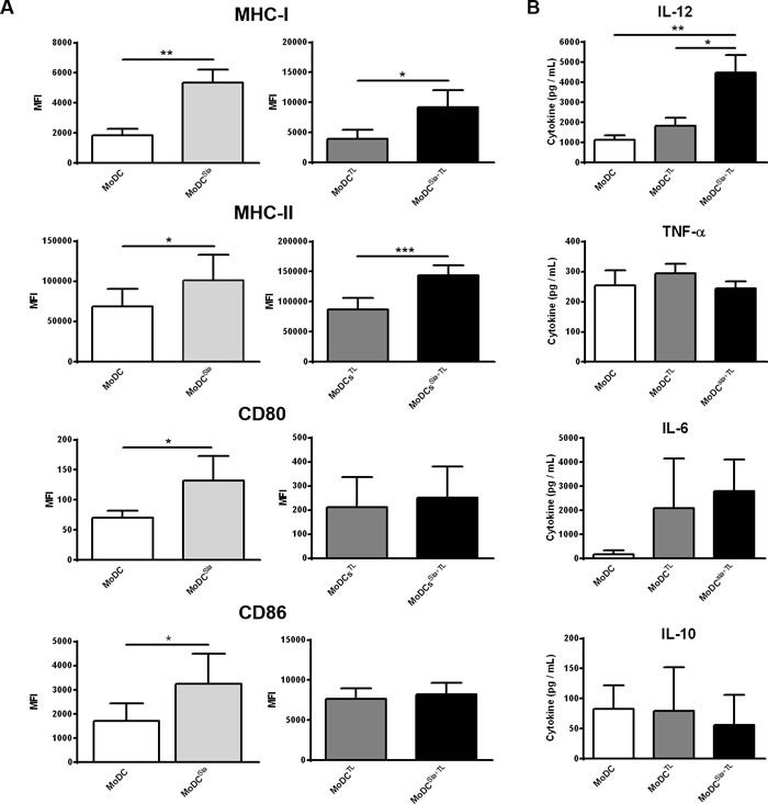
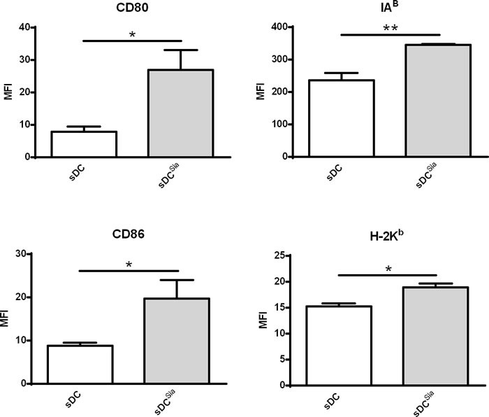
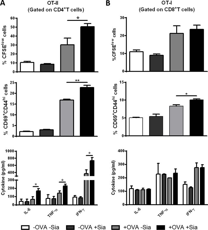
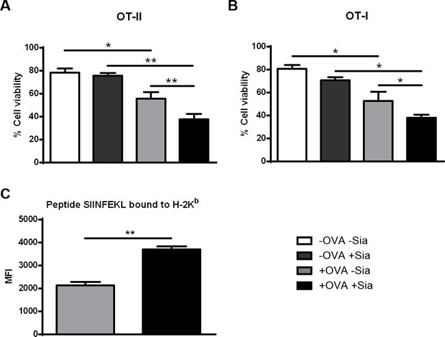
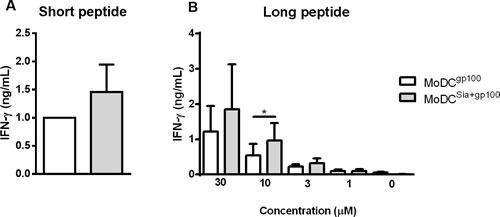
Dendritic cells (DCs) hold promise for anti-cancer immunotherapy. However, clinically, their efficiency is limited and novel strategies to improve DC-mediated anti-tumor responses are needed. Human DCs display high content of sialic acids, which inhibits their maturation and co-stimulation capacity. Here, we aimed to understand whether exogenous desialylation of DCs improves their anti-tumor immunity. Compared to fully sialylated DCs, desialylated human DCs loaded with tumor-antigens showed enhanced ability to induce autologous T cells to proliferate, to secrete Th1 cytokines, and to specifically induce tumor cell apoptosis. Desialylated DCs showed an increased expression of MHC-I and -II, co-stimulatory molecules and an augmented secretion of IL-12. Desialylated HLA-A*02:01 DCs pulsed with gp100 peptides displayed enhanced peptide presentation through MHC-I, resulting in higher activation ofgp100280-288 specific CD8+ cytotoxic T cells. Desialylated murine DCs also exhibited increased MHC and co-stimulatory molecules and higher antigen cross-presentation via MHC-I. These DCs showed higher ability to activate antigen-specific CD4+ and CD8+ T cells, and to specifically induce tumor cell apoptosis. Collectively, our data demonstrates that desialylation improves DCs' ability to elicit T cell-mediated anti-tumor activity, due to increased MHC-I expression and higher antigen presentation via MHC-I. Sialidase treatment of DCs may represent a technology to improve the efficacy of antigen loaded-DC-based vaccines for anti-cancer immunotherapy.

Keywords: Th1-polarization; anti-tumor immunity; antigen cross-presentation; dendritic cells; sialic acid.


Conflict of interest statement

All authors declare no competing financial interests.

Figures

Figure 1
Figure 1. Desialylation of human MoDCs loaded with whole tumor cell antigens induces T cell activation
MoDCs were first treated with sialidase (Sia), for 1 hour at 37°C or left untreated, followed by loading with MCF-7 lysates (TL), as source of whole tumor cell antigens. MoDCs were then used in co-cultures with autologous T cells for 4-8 days in the presence of of IL-2. A. Desialylated MoDCs show a higher induction of T cell proliferation. T cells were previously labeled with CFSE and the progeny, i.e., the percentage of T cells that proliferated, was estimated by flow cytometry, based on the CFSE dilution. The histograms show representative experiments where T cells were co-cultured with: unloaded MoDCs (panel a), MoDCs loaded with MCF-7 lysates (panel b) and sialidase treated MoDCs loaded with MCF-7 lysates (panel c). Unstimulated T cells (panel d) served as negative control and phytohaemagglutinin (PHA)-stimulated T cells (panel e) as positive control. Tabled values represent the mean percentage of proliferative cells ± SEM of 4 independent measurements. B. T cells primed with desialylated MoDCs show higher secretion of Th1 cytokines. The cytokines secreted into the co-culture supernatants, following T cell stimulation with MoDCs were measured by ELISA (n = 7). Graph values represent the concentration (pg/mL) (average ± SEM). Statistically significant differences were calculated using t-test (*P < 0.05).
Figure 2
Figure 2. Desialylation of human MoDCs loaded with whole tumor cell antigens improves T cell cytotoxicity against tumor cells
MoDCs obtained from HLA-A*02:01 donors were treated with sialidase (Sia), for 1 hour at 37°C or left untreated, and loaded with lysates of the breast cancer cell line MCF-7 (TL). Autologous CD3+ T cells (T) were co-cultured with MoDCs loaded with MCF-7 lysates (MoDCTL), or MoDCs that were sialidase treated and loaded with MCF-7 lysates (MoDCsia+TL). After 3 weeks, CD3+ T cells were co-cultured for 5 hours with MCF-7 cells (Tu) in a ratio of 1 Tu: 10 T cells; non-stimulated T cells were cultured with MCF-7 as control. (A., B.) T cells primed with desialylated human MoDCs loaded with MCF-7 cell lysates induce higher tumor cell apoptosis. Tumor cell death was evaluated by flow cytometry by assessing both the intensity of GFP reporter (A) and Annexin V and 7-AAD reactivity. (B) Values represent the percentage ± SEM of viable tumor cells based on the maintenance of GFP fluorescence (GFP+) in 4 independent assays (graph A) and the percentage ± SEM of apoptotic tumor cells assessed by staining with Annexin-V and 7-AAD, in 3 independent measurements (graph B). Statistically significant differences were calculated using t-test (*P < 0.05). C. T cells primed with desialylated MoDCs show higher degranulation. The degranulation of cytotoxic T cells against tumor cells was determined based on the CD107a expression. Representative Histogram is a representative experiment of 4 independent assays.
Figure 3
Figure 3. Desialylated MoDCs show higher levels of maturation and expression of pro-inflammatory cytokines
A. Desialylated MoDCs show a higher maturation phenotype than fully sialylated MoDCs. The expression of several maturation markers was evaluated by flow cytometry. MoDCs were first treated with sialidase (Sia) for 1h at 37°C and then loaded (A, right panel) or not (A, left panel) with MCF-7 lysates (TL), for 3h at 37°C, as described in Materials and methods. Graph values represent the mean fluorescence intensity (MFI) (average ± SEM) of at least 3 independent assays. Statistically significant differences were calculated using t-test (*P < 0.05, **P < 0.01 and ***P < 0.0001) and refers to the difference between untreated and sialidase treated conditions. B. Sialidase treatment of MoDCs elicits IL-12 secretion. The cytokines secreted by MoDCs following sialidase treatment were measured by ELISA. Data represent the cytokine concentration (pg/mL) (average ± SEM) of 6 independent experiments. Statistically significant differences were calculated using t-test (*P < 0.05 and **P < 0.01) and refers to the difference between untreated and sialidase treated conditions.
Figure 4
Figure 4. Sialidase treatment of murine sDCs induces their maturation
Murine sDCs were treated with sialidase or left untreated and the maturation markers evaluated by flow cytometry. Statistical significance (*P < 0.05 or **P < 0.001) refers to the difference between fully sialylated and desialylated sDCs, calculated using t-test. Values represent MFI (average ± SEM) of at least 6 independent assays.
Figure 5
Figure 5. Sialidase treatment of murine sDCs induces OVA-specific CD4+ and CD8+ T cell proliferation and activation
Murine sDCs were pulsed with OVA for 4h and treated or not with sialidase for 1h. A. Desialylated sDCs strongly induce proliferation, activation and differentiation of CD4 + T cells into a Th1 effector phenotype. Splenocytes from OT-II mice were isolated and co-cultured with sDCs for 72h in a ratio of 1:2 (DCs:splenocytes). After 72 h, the percentage of proliferating and activated CD4+ T cells was determined by flow cytometry using the CFSE dilution assay and evaluating the expression of CD69 and CD44, respectively (gating on CD4+ T cells). B. Desialylated sDCs induce activation of CD8 + T cells. Splenocytes from OT-I mice were isolated and co-cultured with sDCs for 72h in a ratio of 1:2 (sDCs:splenocytes) The proliferation of CD8+ T cells was determined by CFSE dilution and the activation determined based on the expression of CD69 and CD44 (gating on CD8+ T cell). The secretion of IFN-γ, IL-6, and TNF-α into the supernatants of co-cultures was evaluated by ELISA. Graph values represent the percentage of CFSElow cells, percentage of CD69+CD44high cells and cytokine concentration (pg/mL) (average ± SEM) from 3 independent assays. Statistical significance (*P < 0.05 or **P < 0.001), calculated using t-test, refers to the difference between fully sialylated and desialylated sDCs.
Figure 6
Figure 6. Sialidase treatment of murine sDCs loaded with OVA improves T cell cytotoxicity against tumor cells
Induction of cytolysis of tumor cells was tested by co-culturing untreated (grey light bars) or sialidase treated (black bars) OVA-pulsed DCs and splenocytes from either OT-II (A.) or OT-I (B.) splenocytes for 72h. After, primed T cells and OVA-expressing B16 mouse melanoma cells (B16-OVA) were co-cultured for 24h. B16-OVA tumor cell death was assessed by staining with Annexin-V and 7-AAD and analysed by flow cytometry. Graph values represent the percentage ± SEM of viable tumor cells of 2 independent assays. Statistical significance (*P < 0.05 or **P < 0.001) refers to the difference between fully sialylated and desialylated sDCs (n = 2). (C.) Desialylated sDCs show improved presentation of endocytosed ovalbumin-derived peptide SIINFEKL, bound to MHC class I. Murine splenic DCs were first incubated with OVA and treated (black bars) or not (grey bars) with sialidase. After these treatments, the presentation of the ovalbumin-derived peptide SIINFEKL bound to H-2Kb (murine MHC class I) epitopes was analysed by flow cytometry, using the 25-D1.16 monoclonal mAb. Graphs show the MFI ± SEM of 2 independent assays. Statistical significance (**P < 0.001) refers to the difference between fully sialylated and desialylated sDCs and it was determined using t-test.
Figure 7
Figure 7. Sialidase treatment of human MoDCs improves antigen cross-presentation via MHC-I to CD8+ T cells
Gp100-specific CD8+ T cells were co-cultured overnight with gp100-loaded MoDCs, that have been previously treated or not with sialidase (MoDCsia+gp100 and MoDCgp100, respectively). To evaluate the activation of CD8+ T cells, the secretion of IFN-γ cytokine was measured by ELISA. A. Short gp100 peptides or B. different concentrations of the long gp100 peptide were used to load MoDCs. Graph values are normalized to the condition of MoDCgp100 with the short peptide. Data refers to the IFN-γ secretion (ng/mL) (average ± SEM) of 3 independent experiments. Statistical significance (*P < 0.05) refers to the difference between fully sialylated and desialylated gp100-loaded MoDCs.

References

    1. Strioga MM, Felzmann T, Powell DJ, Ostapenko V, Dobrovolskiene NT, Matuskova M, Michalek J, Schijns VE. Therapeutic dendritic cell-based cancer vaccines: the state of the art. Crit Rev Immunol. 2013;33:489–547. - PubMed
    1. Sabado RL, Bhardwaj N. Directing dendritic cell immunotherapy towards successful cancer treatment. Immunotherapy. 2010;2:37–56. - PMC - PubMed
    1. Anguille S, Smits EL, Lion E, van Tendeloo VF, Berneman ZN. Clinical use of dendritic cells for cancer therapy. Lancet Oncol. 2014;15:e257–267. - PubMed
    1. Bhargava A, Mishra D, Banerjee S, Mishra PK. Dendritic cell engineering for tumor immunotherapy: from biology to clinical translation. Immunotherapy. 2012;4:703–718. - PubMed
    1. Dawson N. Immunotherapeutic Approaches in Prostate Cancer: PROVENGE. Clinical advances in hematology & oncology. 2010;8:419–421. - PubMed

MeSH terms