. 2016 May 20;12(5):e1005659.
doi: 10.1371/journal.ppat.1005659. eCollection 2016 May.

Nipah Virus C Protein Recruits Tsg101 to Promote the Efficient Release of Virus in an ESCRT-Dependent Pathway

Affiliations

Nipah Virus C Protein Recruits Tsg101 to Promote the Efficient Release of Virus in an ESCRT-Dependent Pathway

Arnold Park et al. PLoS Pathog. .

Abstract

The budding of Nipah virus, a deadly member of the Henipavirus genus within the Paramyxoviridae, has been thought to be independent of the host ESCRT pathway, which is critical for the budding of many enveloped viruses. This conclusion was based on the budding properties of the virus matrix protein in the absence of other virus components. Here, we find that the virus C protein, which was previously investigated for its role in antagonism of innate immunity, recruits the ESCRT pathway to promote efficient virus release. Inhibition of ESCRT or depletion of the ESCRT factor Tsg101 abrogates the C enhancement of matrix budding and impairs live Nipah virus release. Further, despite the low sequence homology of the C proteins of known henipaviruses, they all enhance the budding of their cognate matrix proteins, suggesting a conserved and previously unknown function for the henipavirus C proteins.


Conflict of interest statement

The authors have declared that no competing interests exist.

Figures

Fig 1
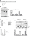
Fig 1. Nipah virus budding is ESCRT-dependent.
(A) Wild-type (WT) and dominant-negative (DN) Vps4A-inducible 293 cells were infected with Gaussia luciferase-expressing NiV at a multiplicity of infection (MOI) of 2. Cells were induced with doxycycline (dox) post-infection, and viral titers in the supernatant were determined at the indicated time points. Error bars represent standard deviations for 3 replicates. Induction of DN Vps4A significantly inhibits the release of NiV at 24 hours post-infection (hpi). ***, p<0.001, two-way ANOVA followed by Bonferroni posttests. (B) Induction of WT or DN Vps4A did not affect Gaussia luciferase (Gluc) production, indicating unimpaired virus protein production. ns, not significant, two-way ANOVA followed by Bonferroni posttests. (C) Vps4A-inducible 293 cells were infected with wild-type NiV at MOI 2, and Vps4A (WT vs. DN) was induced (+Dox) or not (-Dox) immediately after infection as in Fig 1A. At 24 hpi, cell lysates (CL) were collected, and virions were ultracentrifuged through 20% sucrose and resuspended in Laemmli SDS sample buffer for Western analysis. The 0 hpi time point represents the last wash after removal of the inoculum to determine the background level of residual virus. Induction of DN Vps4A clearly resulted in less virions (NiV) released into the supernatant.]
Fig 2
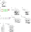
Fig 2. NiV-M budding is enhanced by NiV-C in an ESCRT-dependent manner.
(A) A schematic of the NiV genome is shown, with the NiV genes arrayed between the genomic termini, the 3’ leader (3’le) and 5’ trailer (5’tr). NiV-V and -W are produced by the stochastic inclusion of additional G nucleotides in the P mRNA via polymerase stuttering (as indicated in the schematic), thus creating a frameshift at that juncture, while NiV-C is produced off an alternate reading frame. (B) NiV-M was co-transfected with vector only or with HA-tagged NiV-derived gene products into 293T cells, and relative virus-like particle budding was determined as described in Materials and Methods. COX IV is shown as the cell lysate loading control. Error bars represent standard deviations for 3 independent experiments. Only NiV-C had a significant effect on M budding relative to the vector control. ***, p<0.001, one-way ANOVA followed by Bonferroni posttests. (C) CCL-81 Vero cells were infected with Gluc-expressing WT or NiV-C knockout NiV at MOI 2. Virus titers were determined from supernatant collected at 24 hpi. Error bars represent standard deviations for 3 replicates. **, p<0.01, 2-tailed Student’s t-test. (D) At 24 hpi, cell lysates (CL) were collected, and virions were ultracentrifuged through 20% sucrose and resuspended in Laemmli SDS sample buffer for Western analysis. Normalized relative budding values are shown below the blot. (E) Knockout of NiV-C did not affect Gluc production, indicating unimpaired protein production. ns, not significant, two-way ANOVA followed by Bonferroni posttests. (F) Vps4A-inducible 293 cells were transfected with NiV-M and with or without NiV-C-HA as indicated. At 4 hours post-transfection, media was changed with or without doxycycline. Error bars represent standard deviations for 3 independent experiments. **, p<0.01, one-way ANOVA followed by Bonferroni posttests. Only NiV-C enhancement of NiV-M-mediated VLP budding was significantly affected by induction of DN Vps4A.
Fig 3
Fig 3. NiV-C interacts with NiV-M to promote budding.
(A) NiV-C-HA and NiV-M were transfected as indicated in 293T cells, and lysates were immunoprecipitated with anti-NiV-M as described in Materials and Methods. NiV-C was detected with anti-HA. (B) NiV-C-HA and NiV-M were transfected as indicated in 293T cells. NiV-C was detected with anti-HA. NiV-C is only detected in VLPs in the presence of NiV-M VLP budding. (C) NiV-M and NiV-C-HA were co-transfected in 293T cells and detected by confocal microscopy as described in Materials and Methods. DAPI (blue) illustrates the nuclei. A representative Z-slice is shown.
Fig 4
Fig 4. C enhancement requires a cluster of residues conserved between NiV-C and host factor Vps28.
(A) A structural homology modeling search of NiV-C in the Phyre2 server (Imperial College, London) revealed an alignment with the C-terminal domain (CTD) of Vps28 of X. laevis (Xl-Vps28). Based on the alignment, we defined a N-terminal domain (NTD), middle domain (MiD), and C-terminal domain (CTD) for NiV-C. The presence of a double glycine (GG) immediately following NiV-C-MiD is suggestive of a linker between domains. The sequence alignment was created in Clustal Omega, with illustration via BOXSHADE, with conserved (black) and similar (grey) residues highlighted. The CTD of human Vps28 (Hs-Vps28) is included in the alignment for comparison. A nearly identical stretch of 7 amino acids between NiV-C and Hs-Vps28 is boxed in orange. (B) Left, the X. laevis Vps28-CTD structure (dark blue) is shown, with the stretch of 7 amino acids indicated in the sequence alignment in Fig 4A highlighted in orange in their homologous positions [26] (PDB 2J9W, displayed in Chimera). The N- to C-terminal helices of the four-helix bundle are labeled as α1–4. Right and boxed, rotated view of S. cerevisiae Vps28-CTD (Sc-Vps28-CTD, light blue) and Vps36 Npl4 zinc-finger N-terminal domain (Sc-Vps36-NZF-N, magenta), shown in their known structural interaction [26] (PDB 2J9U). The same stretch of 7 amino acids is also highlighted here in orange in their homologous positions (residues 203–209 in Sc-Vps28) in the Sc-Vps28-CTD structure. Residues I203, I206, and V207 within this stretch are functionally important in the yeast Vps28-Vps36 interface [26] and their side chains are illustrated. Black and green highlight in the Sc-Vps28-CTD structure illustrate residues discussed in Fig 4C (see following). (C) Alanine mutagenesis in full length NiV-C-HA was performed as shown. The positions of P88 and M110 in their homologous positions in Sc-Vps28-CTD are shown in black and green highlight (D194 in black and L212 in green, respectively) in Fig 4B. P88 is within the loop between helices 2 and 3, which differs by 2 residues in length between metazoan and yeast Vps28. Due to differing alignments in this region, the closest residue in yeast Vps28 (D194) in most considered alignments was chosen for comparison [–28]. Mutation of W103 and L104 within the highlighted stretch of residues selectively abrogates the ability of NiV-C to enhance NiV-M budding. Error bars represent standard deviations for 3 independent experiments. ***, p<0.001, one-way ANOVA followed by Bonferroni posttests.
Fig 5
Fig 5. NiV-C has a direct interaction with Tsg101.
(A) HA-tagged NiV-C co-immunoprecipitates endogenous Tsg101 from 293T cell lysates, as does HA-tagged Vps28, a known cellular interacting partner of Tsg101. (B) NiV-C was truncated as indicated, with all constructs retaining a C-terminal HA tag. (C) Loss of the CTD of NiV-C specifically leads to loss of pulldown of Tsg101. (D) A fusion of EGFP and NiV-C-CTD (HA-EGFP-C-CTD) pulls down Tsg101, whereas EGFP alone does not. (E) The C terminal domain (CTD) of Tsg101 was defined as in [31]. FLAG-Tsg101 was truncated as indicated. UEV, ubiquitin E2 variant domain; PRO, proline-rich domain; CC, coiled-coil region; and CTD, C-terminal domain. (F) Truncation of the CTD of Tsg101 results in loss of co-immunoprecipitation with NiV-C. (G) A fusion of EGFP and the full CTD (amino acids 303–390) of Tsg101 (FLAG-EGFP-Tsg101-CTD) co-immunoprecipitates with NiV-C, whereas EGFP alone does not. (H) Left panel, Coomassie-stained gel showing recombinant proteins (100 ng each lane) purified from E. coli as described in Materials and Methods. Right panel, Western analysis with repeat of Fig 5G except with purified recombinant proteins, showing that the interaction between NiV-C and Tsg101-CTD is direct.
Fig 6
Fig 6. Tsg101 is required for the NiV-C enhancement of budding and efficient release of live NiV.
(A) NiV-C was minimally truncated at its C-terminus as indicated, with all constructs retaining a C-terminal HA tag. (B) Truncation of only 7 residues (159tr) from NiV-C results in loss of interaction with Tsg101. (C) This truncation mutant no longer enhances NiV-M budding. Error bars represent standard deviations for 3 independent experiments. ns, not significant; ***, p<0.001, one-way ANOVA followed by Bonferroni posttests. (D) Schematic of strategy to insert a destabilization domain (DD) tag onto all endogenous copies of Tsg101, via CRISPR/Cas9-mediated homologous recombination (HR). Exon 1 of Tsg101 in the human genome is shown, with the start codon highlighted in orange. Light blue highlight represents sequence used in the homology arms in the donor constructs. Once integrated into the genome, antibiotic resistance is driven off of the endogenous promoter, with the P2A ribosomal skipping sequence allowing the DD-Tsg101 fusion product to be translated separately. Green arrows indicate position of genotyping primers used for Fig 6E. (E) Genotyping PCR of the single cell-isolated DD-Tsg101 293T clone with complete knock-in (KI), using genome-specific primers (see Fig 6D, green arrows) flanking the homology arms used in the donor constructs. Asterisk represents a background product. The puromycin and blasticidin donors insert 1,182 and 981 bp, respectively, hence the doublet seen in the knock-in PCR product. (F) Knock-in DD-Tsg101 cells (KI) were incubated with 10 uM trimethoprim (TMP) (+ TMP) or DMSO vehicle control (- TMP) for 24h, then collected for comparison with parental 293T cells (WT) by Western. The upward shift in molecular weight for DD-Tsg101 is due to the 18 kDa DD tag fusion. The relative expression of Tsg101 indicated below the blot was normalized to the COX IV loading control. (G) DD-Tsg101 cells were incubated in 10 uM TMP or DMSO vehicle as described in Materials and Methods before transfection with NiV-M and with or without NiV-C as indicated. Destabilization of DD-Tsg101 by removal of TMP abrogates the NiV-C enhancement of NiV-M budding. Error bars represent standard deviations for 3 independent experiments. ns, not significant; **, p<0.01, 2-tailed Student’s t-test. (H) DD-Tsg101 cells in 10 uM TMP or DMSO vehicle were infected with wild-type NiV at MOI 2 or Rift Valley fever virus (RVFV) at MOI 0.2, and supernatant titers were determined at 24 hpi. Error bars represent standard deviations for 3 replicates. ns, not significant; *, p<0.05, 2-tailed Student’s t-test. (I) DD-Tsg101 cells in 10 uM TMP or DMSO vehicle were infected with wild-type NiV at MOI 2 and processed for TEM as described in Materials and Methods. All fields are shown at the same magnification. Right panels, samples were stained with anti-NiV-N and 15 nm colloidal gold. White arrows show examples of immunogold staining.
Fig 7
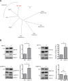
Fig 7. The C proteins of other known henipaviruses also enhance the budding of their cognate matrix proteins.
(A) Phylogeny (based on the relatively conserved nucleocapsid protein, neighbor-joining tree from Clustal Omega, visualized in FigTree) for the henipaviruses as well as the “henipa-like” Mojiang virus (MojPV) and representative paramyxoviruses from each of the other major genera within the Paramyxovirinae subfamily: Measles virus (MeV, Genbank AB016162.1), Newcastle disease virus (NDV, Genbank AF309418.1), Mumps virus (MuV, AB040874.1), and human parainfluenza virus 1 (HPIV-1, Genbank AF457102.1). NiV and HeV, as the prototypic members of Henipavirus, are highlighted in red. (B) HA-tagged matrix and C proteins were transfected in 293T cells as indicated. All the C proteins enhance the budding of their cognate M proteins, with the exception of “henipa-like” MojPV. Error bars represent standard deviations for 4 independent experiments. ns, not significant; *, p<0.05; **, p<0.01, 2-tailed Student’s t-test.
Fig 8
Fig 8. Model of NiV-C interactions with ESCRT.
Vps28 acts as an adaptor between ESCRT-I and ESCRT-II. The NTD of Vps28 interacts with ESCRT-I via Tsg101-CTD, while the CTD of Vps28 interacts with ESCRT-II possibly via hVps36. The CTD of NiV-C also interacts with Tsg101-CTD, while the potential interaction of NiV-C-MiD with ESCRT-II or other downstream ESCRT factors remains to be defined.

Similar articles

Cited by

References

    1. Henne WM, Buchkovich NJ, Emr SD (2011) The ESCRT pathway. Dev Cell 21: 77–91. 10.1016/j.devcel.2011.05.015 - DOI - PubMed
    1. Schmidt O, Teis D (2012) The ESCRT machinery. Curr Biol 22: R116–120. 10.1016/j.cub.2012.01.028 - DOI - PMC - PubMed
    1. Votteler J, Sundquist WI (2013) Virus budding and the ESCRT pathway. Cell Host Microbe 14: 232–241. 10.1016/j.chom.2013.08.012 - DOI - PMC - PubMed
    1. Bieniasz PD (2009) The cell biology of HIV-1 virion genesis. Cell Host Microbe 5: 550–558. 10.1016/j.chom.2009.05.015 - DOI - PMC - PubMed
    1. Demirov DG, Freed EO (2004) Retrovirus budding. Virus Res 106: 87–102. - PubMed

Publication types

MeSH terms