. 2016 Sep 29;24(9):1570-80.
doi: 10.1038/mt.2016.105. Epub 2016 May 16.

High Efficiency CRISPR/Cas9-mediated Gene Editing in Primary Human T-cells Using Mutant Adenoviral E4orf6/E1b55k "Helper" Proteins

Affiliations

High Efficiency CRISPR/Cas9-mediated Gene Editing in Primary Human T-cells Using Mutant Adenoviral E4orf6/E1b55k "Helper" Proteins

Kamila S Gwiazda et al. Mol Ther. .

Abstract

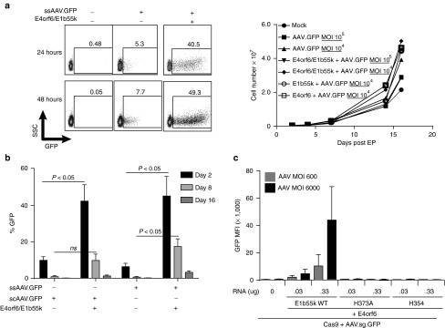
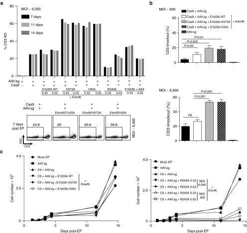
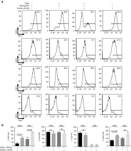
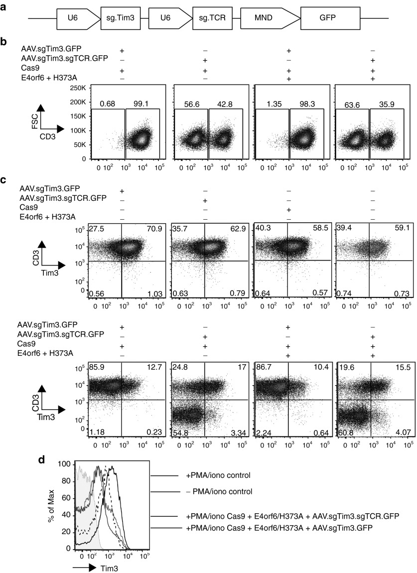
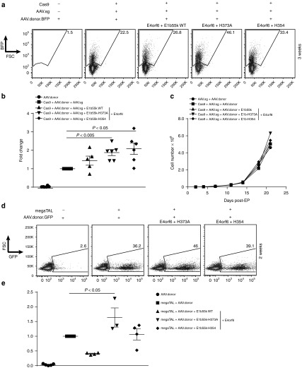
Many future therapeutic applications of Clustered Regularly Interspaced Short Palindromic Repeats (CRISPR)/Cas9 and related RNA-guided nucleases are likely to require their use to promote gene targeting, thus necessitating development of methods that provide for delivery of three components-Cas9, guide RNAs and recombination templates-to primary cells rendered proficient for homology-directed repair. Here, we demonstrate an electroporation/transduction codelivery method that utilizes mRNA to express both Cas9 and mutant adenoviral E4orf6 and E1b55k helper proteins in association with adeno-associated virus (AAV) vectors expressing guide RNAs and recombination templates. By transiently enhancing target cell permissiveness to AAV transduction and gene editing efficiency, this novel approach promotes efficient gene disruption and/or gene targeting at multiple loci in primary human T-cells, illustrating its broad potential for application in translational gene editing.


Figures

Figure 1
Figure 1
Enhanced AAV-mediated gene expression in primary human T-cells using adenoviral E4orf6/E1b55k proteins to relieve postentry AAV restriction mechanisms. (a) Relief of postentry restriction of AAV-mediated gene expression. Left panel: AAV-mediated GFP expression following relief of postentry AAV restriction by E4orf6/E1b55k. Primary CD4+ T-cells were electroporated with mRNA encoding adenoviral serotype 5 E4orf6/E1b55k (0.33 μg each), rested for 2–4 hours, then transduced with AAV driving GFP expression. Cells were placed in culture for the indicated periods of time, following which the cells were collected and analyzed for GFP expression by flow cytometry. Right panel: Expansion of cell populations following the indicated exposure to E4orf6/E1b55k mRNA transfection and AAV transduction, following the same protocol as described in the left panel. (b) Comparison of effect of E4orf6/E1b55k proteins on self-complementary and single-stranded AAV6-mediated gene expression. Primary CD4+ T-cells were electroporated with mRNA encoding E4orf6/E1b55k proteins (0.33 μg each), rested for 2–4 hours, and transduced with either single-stranded or self-complementary AAV6 driving GFP expression (MOI of both viruses: 2 × 104). Cells were placed in culture for the indicated periods of time, following which the cells were collected and analyzed for GFP expression by flow cytometry. (c) E1b55k Mre11/Rad51/NBS1 (MRN) degradation mutants demonstrate requirement for MRN inactivation for complete relief of postentry restriction on AAV-mediated expression. Primary CD4+ T-cells were electroporated with mRNA encoding Cas9-2A-mCherry (1 µg) along with E4orf6/E1b55k (Wild type) proteins or the indicated E1b55k mutants at the indicated RNA doses, rested for 2–4 hours, and transduced with ssAAV driving both TCRα guide and GFP expression. E4orf6 mRNA dose was the same as each E1b55k RNA dose for each indicated point. Cells were placed in culture for two days, following which the cells were collected and analyzed for GFP MFI by flow cytometry. See also Supplementary Figure S2 demonstrating a 2–3 fold increase in GFP expression catalyzed by the E4orf6/E1b55k H373A mutant complex that is not visible on the same scale as GFP expression catalyzed by the WT E4orf6/E1b55k complex.
Figure 2
Figure 2
Enhanced CRISPR-mediated gene knockout in primary human T-cells through use of adenoviral E4orf6/E1b55k proteins. (a) E1b55k mutants enhance gene knockout achieved using mRNA/AAV delivery of CRISPR components. Representative experiment indicating primary CD4+ T-cells electroporated with mRNA encoding Cas9-T2A-mCherry, (1 μg), E4orf6/E1b55k (Wild Type), or the indicated mutants (at 0.33 μg or 0.03 μg each), rested for 2–4 hours, and transduced with AAV driving TCRα guide expression. Cells were placed in culture, following which the cells were collected and analyzed for CD3 expression by flow cytometry at the indicated time-points following EP/transduction. Upper panel—quantification of CD3 knockout. Bottom panel—representative flow plots from a subset of the experiment at 7 days post EP/transduction. (b) E4orf6/E1b55k MRN-inactivation deficient mutants enhance CRISPR-mediated knockout. Quantification of n = 3–4 independent experiments at two different AAV MOIs indicating that both E1b55k H373A and H354 mutants significantly increase CRISPR-mediated TCRα knockout, quantified by CD3 staining at 7 days post EP/transduction. Primary human CD4+ or CD3+ T-cells were used. (c) T-cells edited using mRNA/AAV delivery exhibit normal expansion kinetics. Primary CD4+ T-cells were electroporated with mRNA encoding Cas9-T2A-mCherry (1 μg) proteins along with Wild Type E4orf6/E1b55k proteins or the indicated E1b55k mutants, rested for 2–4 hours, and transduced with AAV driving TCRα guide expression. Cells were placed in culture for the indicated periods of time, during which aliquots of the cells were collected and counted for quantification of cell expansion. Left panel: low dose (0.03 μg) of E4orf6/E1b55k mutants were electroporated with high dose of AAV (MOI ~ 6000). Right panel: both low and high dose (0.33 μg) E4orf6/E1b55k-R240A RNA, and low and high dose AAV, were electroporated and transduced.
Figure 3
Figure 3
Implementation of CRISPR/Cas9 with mRNA/AAV delivery is able to achieve efficient knockout at multiple genomic sites. (a) Primary human CD3+ T-cells were electroporated using the MaxCyte GT system with mRNA encoding Cas9-2A-mCherry proteins (1 µg) along with E4orf6/E1b55k-H373A proteins (0.03 μg each), rested for 2–4 hours, and transduced with AAV driving guide expression against the indicated surface proteins. Cells were placed in culture and allowed to expand; 9–12 days following initial stimulation, cells were restimulated using Dynal CD3/CD28 beads for 48 hours, following which the cells were collected and analyzed for expression of the indicated surface proteins by flow cytometry and assessed for knockout by amplicon sequencing. Representative flow cytometry plots indicating that T-cells edited using mRNA/AAV codelivery exhibit loss of targeted surface checkpoint proteins. Data represent n = 3–5 independent editing experiments. (b) Quantification and summary data from flow cytometry analysis of gene knockout and sequencing analysis of amplicons from genomic target sites. Analysis of knockout by flow cytometry is assessed by percent loss of induction of surface protein expression. Data represent n = 3–5 independent editing experiments. ND, not determined.
Figure 4
Figure 4
Cas9 mRNA/AAV guide delivery is able to achieve efficient CRISPR-mediated multiplex knockout in primary human T-cells with E4orf6/E1b55k H373A expression. Primary human CD3+ T-cells were electroporated using the MaxCyte GT system with mRNA encoding Cas9-2A-mCherry proteins (1 µg) along with E4orf6/E1b55k-H373A proteins (0.03 μg each), rested for 2–4 hours, and transduced with AAVs driving guide expression against Tim3 and TCRα, as well as GFP expression to track transduction efficiency. Cells were placed in culture and allowed to expand; 7 days following EP/transduction, cells were assessed for TCRα knockout by CD3 stain. Three weeks following initial stimulation, cells were restimulated using PMA/ionomycin for 3–4 hours and allowed to recover for 48 hours, following which the cells were collected and analyzed for expression of Tim3 by flow cytometry. (a) Schematic of the multiplex AAV vector expressing guide RNAs against Tim3 and TCRα, with individual U6 promoters. (b) Representative flow cytometry analysis of TCRα knockout by CD3 staining, 7 days after EP/transduction. (c) Representative flow cytometry analysis of TCRα knockout and Tim3 knockout, 3 weeks after initial stimulation. To upregulate Tim3 surface expression independently of TCR, cells were stimulated using PMA/ionomycin (10 ng/ml and 1 µg/ml, respectively), for 3–4 hours and rested for 48 hours. (d) Upregulation of Tim3 with and without PMA/ionomycin stimulation. Shown are control cells (AAV treatment only) with and without PMA/ionomycin treatment, and stimulated cells with Tim3 knockout compared with stimulated cells missing both Tim3 and TCRα. There were no differences between cells proficient and deficient in TCRα signaling, suggesting that PMA/ionomycinn stimulation is independent of this pathway. In shaded gray are unstained cells.
Figure 5
Figure 5
Enhanced targeted knock-in using E1b55k mutant proteins. E4orf6/E1b55k H373A and H354 mutants enhance CRISPR- and megaTAL-mediated recombination achieved with mRNA/AAV codelivery. (a) Primary CD4+ or CD3+ T-cells were electroporated using the MaxCyte GT or the Neon systems with mRNA encoding Cas9-2A-mCherry proteins (1 μg) along with Wild Type E4orf6/E1b55k proteins or the indicated E1b55k mutants (0.03 μg each), rested for 2–4 hours, and transduced with separate AAVs driving CCR5 guide expression and a targeting template for the CCR5 locus (See Supplementary Figure S7). Cells were placed in culture, following which the cells were collected and analyzed for BFP expression by flow cytometry. Shown are representative flow plots from the indicated manipulations at 3 weeks post EP/transduction, from n = 5–6 independent experiments. (b) Quantification of n = 5–6 independent experiments of the fold change in knock-in frequency (BFP+ cells) over baseline (Cas9+ guide + donor) at 3 weeks post EP/transduction. (c) T-cells edited via mRNA/AAV CRISPR-mediated recombination exhibit normal expansion kinetics. Primary T-cells were electroporated with mRNA encoding control or Cas9-2A-mCherry proteins (1 µg) along with Wild Type E4orf6/E1b55k and the E1b55k mutants (0.03 μg each), rested for 2–4 hours, and transduced with AAV driving CCR5 guide expression as well as AAV CCR5 BFP template. Cells were placed in culture for the indicated periods of time, following which the cells were collected and counted for quantification of cell expansion. (d) Primary CD4+ or CD3+ T-cells were electroporated using the MaxCyte GT or the Neon systems with mRNA encoding CCR5 megaTAL (0.5–1 μg) along with Wild Type E4 or f6/E1b55k proteins or the indicated E1b55k mutants (0.03 μg each), rested for 2–4 hours, and transduced with a GFP targeting template for the CCR5 locus. Cells were placed in culture, following which the cells were collected and analyzed for GFP expression by flow cytometry. Shown are representative flow plots from the indicated manipulations, at 2 weeks post EP/transduction, from n = 3–5 independent experiments. (e) Quantification of n = 3–5 independent experiments of the fold change in knock-in frequency (GFP+ cells) over baseline (megaTAL + donor) at 2 weeks post EP/transduction. We found the fold change in HDR was significantly increased using E4orf6/E1b55k-H373A only.

Similar articles

Cited by

References

    1. Gori, JL, Hsu, PD, Maeder, ML, Shen, S, Welstead, GG and Bumcrot, D (2015). Delivery and specificity of CRISPR-Cas9 genome editing technologies for human gene therapy. Hum Gene Ther 26: 443–451. - PubMed
    1. Jiang, W and Marraffini, LA (2015). CRISPR-Cas: New tools for genetic manipulations from bacterial immunity systems. Annu Rev Microbiol 69: 209–228. - PubMed
    1. Xu, J, Ren, X, Sun, J, Wang, X, Qiao, HH, Xu, BW et al. (2015). A toolkit of CRISPR-based genome editing systems in Drosophila. J Genet Genomics 42: 141–149. - PubMed
    1. Sánchez-Rivera, FJ and Jacks, T (2015). Applications of the CRISPR-Cas9 system in cancer biology. Nat Rev Cancer 15: 387–395. - PMC - PubMed
    1. Vasileva, EA, Shuvalov, OU, Garabadgiu, AV, Melino, G and Barlev, NA (2015). Genome-editing tools for stem cell biology. Cell Death Dis 6: e1831. - PMC - PubMed

Publication types