. 2016 Aug;24(8):1388-94.
doi: 10.1038/mt.2016.107. Epub 2016 May 20.

CRISPR Repair Reveals Causative Mutation in a Preclinical Model of Retinitis Pigmentosa

Affiliations

CRISPR Repair Reveals Causative Mutation in a Preclinical Model of Retinitis Pigmentosa

Wen-Hsuan Wu et al. Mol Ther. 2016 Aug.

Abstract

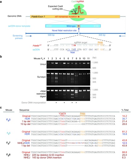
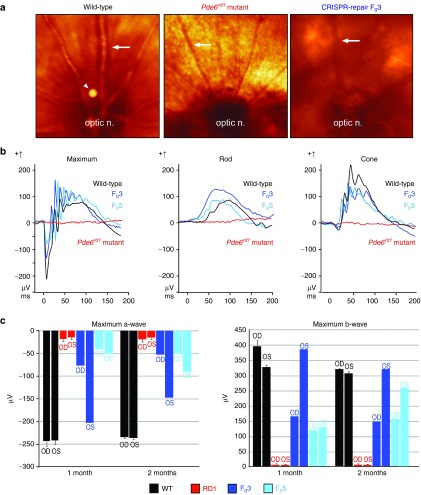
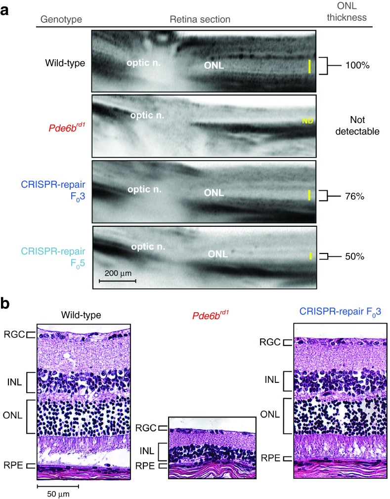
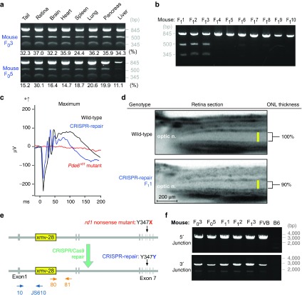
Massive parallel sequencing enables identification of numerous genetic variants in mutant organisms, but determining pathogenicity of any one mutation can be daunting. The most commonly studied preclinical model of retinitis pigmentosa called the "rodless" (rd1) mouse is homozygous for two mutations: a nonsense point mutation (Y347X) and an intronic insertion of a leukemia virus (Xmv-28). Distinguishing which mutation causes retinal degeneration is still under debate nearly a century after the discovery of this model organism. Here, we performed gene editing using the CRISPR/Cas9 system and demonstrated that the Y347X mutation is the causative variant of disease. Genome editing in the first generation produced animals that were mosaic for the corrected allele but still showed neurofunction preservation despite low repair frequencies. Furthermore, second-generation CRISPR-repaired mice showed an even more robust rescue and amelioration of the disease. This predicts excellent outcomes for gene editing in diseased human tissue, as Pde6b, the mutated gene in rd1 mice, has an orthologous intron-exon relationship comparable with the human PDE6B gene. Not only do these findings resolve the debate surrounding the source of neurodegeneration in the rd1 model, but they also provide the first example of homology-directed recombination-mediated gene correction in the visual system.


Figures

Figure 1
Figure 1
CRISPR/Cas9-mediated repair of Y347X was achieved in a mosaic fashion in 2 of 11 Founders (F0). (a) sgRNA targets exon 7 of the Pde6b locus (yellow box, top). Donor template (blue box, middle) encoding the wild-type allele corrects Y347X through homology-directed recombination. Donor template was made resistant to Cas9 by two modifications: (i) NdeI site and (ii) four wobble base pairs. Primers amplify an 845-bp fragment. (b) Surveyor assay reveals that 7 of 11 F0 mice have CRISPR-induced DSBs. NdeI digestion detects correction of Y347X in F03 and F05. In F08 and F07, INDELs were incorporated alongside the ssODN. (c) Pde6b was repaired in 35.7% of F03 and 18.8% of F05 somatic cells. Allelic heterogeneity due to nonhomologous end joining was apparent as small deletions (1–10 bp) in F03 and F07, and large INDELs in F08, were detected. (Red letters: Y347X mutation; blue letters: wobble nucleotides carried by donor template; yellow letters: PAM sequence; brown letters: INDELs).
Figure 2
Figure 2
CRISPR-mediated repair of Y347X in Pde6b restored neurophysiology. (a) Uniform coloration of the fundus of an age-matched wild-type (WT) control. “Salt and pepper” retinopathy in the dorsal fundus of the rd1 mutant as well as vascular attenuation were observed (ON, optic nerve; arrows point to vessels; arrow head points out an imaging artifact). CRISPR-corrected F03 displays a fundus similar to that of WT, with preservation of vasculature. (b) Electroretinogram (ERG) (μV) revealed neurophysiological rescue in 1-month-old gene-corrected mice. Rd1 mice (red) had extinguished mixed maximum rod–cone, rod-specific, and cone ERG responses. In CRISPR-repaired F03 (dark blue) and F05 (light blue) mice, maximum, rod, and cone ERG responses were comparable with WT (black) and significantly heightened compared with the rd1 mutant. (c) Greater magnitude represents improved neurophysiology. Maximum a- and b-wave values obtained at 1 and 2 months in rd1 (red), F03 (dark blue), F05 (light blue), and WT (black) mice. Rd1 consistently displayed significantly diminished a-waves, which are photoreceptor mediated, and b-waves, which are inner retina mediated. F03 OS originally registered as nearly equivalent to the WT, but gradually decreased over time. a- and b-wave magnitudes in F03 OD and F05 OS/OD were poorer than that of WT but still greater than that of the rd1 mutant.
Figure 3
Figure 3
CRISPR-mediated repair of Y347X in Pde6brd1 restored retinal structure. (a) Optical coherence tomography data at 3 months from wild-type (WT), rd1, F03, and F05 mice reflected partial preservation of retinal layers in the CRISPR-rescued mice compared with the rd1 mutant. The outer nuclear layer (ONL) was not detectable in rd1. Yellow bar indicates the ONL thickness, which was normalized to WT and measured to be 76% and 50% in F03 and F05, respectively. (b) Hematoxylin–eosin-stained retinal section from WT, rd1, and F03 mice at 3 months revealed that retinal layers were preserved in F03 and were comparable with that of WT, whereas the rd1 mutant layers had collapsed and loss the photoreceptor (ONL) layer. INL, inner nuclear layer; RGC, retinal ganglion cell; RPE, retinal-pigmented epithelium.
Figure 4
Figure 4
Xmv-28 viral insertion was retained but benign in gene-repaired mice. (a) Restriction fragment polymorphism (RFLP) revealed CRISPR-mediated correction in 24–37% of cells comprising organs in F03, and 11–30% in F05. Data show that the CRISPR-mediated correction occurred at preimplantation, since repaired and uncorrected alleles were uniformly distributed in endoderm, mesoderm, and ectoderm organ lineages. (b) RFLP demonstrates that F03 backcrossed with the Rd1 mutant transmitted the corrected Y347X allele to 3 of 10 offspring (F11, 2, and 3). (c) Mixed maximal rod–cone electroretinogram (ERG) recordings from F1 at P30. F11 (blue) registered a- and b-wave amplitudes similar to that of the wild-type (WT) control (black); rd1 (red) registered a flat ERG. (d) Optical coherence tomography compared retinal layer thicknesses in WT and F11. The latter's outer nuclear layer (ONL) was 90% of WT thickness, confirming rescue was transmitted to F1. (e) Schematic representation of the Xmv-28 insertion (yellow box). CRISPR repairs the Y347X mutation (red), restoring it to the WT sequence (blue) while leaving the insertion intact. Dark blue and orange arrows represent primers used to screen the 5′ and 3′ ends of the viral insertion. Primer 10 targeted Pde6b exon 1, while JS610 targeted the Xmv-28 env gene, together amplifying the 5′ end of the insertion. Primer 81 targeted intron 1 of Pde6b, while primer 80 targeted the Xmv-28 gag gene, together amplifying the 3′ end of the insertion. (f) PCR confirmed that Xmv-28 insertion was retained in CRISPR/Cas9-repaired rd1 mice. This suggests that the Y347X mutation, rather than the viral insertion, causes the disease phenotype.

References

    1. Pittler, SJ, Keeler, CE, Sidman, RL and Baehr, W (1993). PCR analysis of DNA from 70-year-old sections of rodless retina demonstrates identity with the mouse rd defect. Proc Natl Acad Sci USA 90: 9616–9619. - PMC - PubMed
    1. Bowes, C, Li, T, Danciger, M, Baxter, LC, Applebury, ML and Farber, DB (1990). Retinal degeneration in the rd mouse is caused by a defect in the beta subunit of rod cGMP-phosphodiesterase. Nature 347: 677–680. - PubMed
    1. Bowes, C, Li, T, Frankel, WN, Danciger, M, Coffin, JM, Applebury, ML et al. (1993). Localization of a retroviral element within the rd gene coding for the beta subunit of cGMP phosphodiesterase. Proc Natl Acad Sci USA 90: 2955–2959. - PMC - PubMed
    1. Pittler, SJ and Baehr, W (1991). Identification of a nonsense mutation in the rod photoreceptor cGMP phosphodiesterase beta-subunit gene of the rd mouse. Proc Natl Acad Sci USA 88: 8322–8326. - PMC - PubMed
    1. Keeler, CE (1928). The geotropic reaction of rodless mice in light and in darkness. J Gen Physiol 11: 361–368. - PMC - PubMed

MeSH terms

Substances