. 2016 Jun 21;7(25):38243-38256.
doi: 10.18632/oncotarget.9430.

Drug conjugated nanoparticles activated by cancer cell specific mRNA

Affiliations

Drug conjugated nanoparticles activated by cancer cell specific mRNA

Nathan P Gossai et al. Oncotarget. .

Abstract

We describe a customizable approach to cancer therapy in which a gold nanoparticle (Au-NP) delivers a drug that is selectively activated within the cancer cell by the presence of an mRNA unique to the cancer cell. Fundamental to this approach is the observation that the amount of drug released from the Au-NP is proportional to both the presence and abundance of the cancer cell specific mRNA in a cell. As proof-of-principle, we demonstrate both the efficient delivery and selective release of the multi-kinase inhibitor dasatinib from Au-NPs in leukemia cells with resulting efficacy in vitro and in vivo. Furthermore, these Au-NPs reduce toxicity against hematopoietic stem cells and T-cells. This approach has the potential to improve the therapeutic efficacy of a drug and minimize toxicity while being highly customizable with respect to both the cancer cell specific mRNAs targeted and drugs activated.

Keywords: anti-sense; drug delivery; gold nanoparticles; leukemia; molecularly targeted therapy.


Conflict of interest statement

The authors disclose no potential conflicts of interest.

Figures

Figure 1
Figure 1. Development of an Au-NP based system for selective drug activation in cancer cells mediated by cancer cell specific mRNA
Each gold particle is conjugated to ~150-200 oligonucleotides (red) via a thiol linker. The sequence of the oligonucleotide is complementary (anti-sense) to a mRNA that is either overexpressed in or unique to cancer cells. A shorter, complementary drug-conjugated oligonucleotide (drug-orange; oligonucleotide-green) is annealed to the anti-sense oligonucleotide to generate a drug-DNA Au-NP. After cellular uptake, the targeted mRNA (blue) binds to the complementary DNA sequences linked to the Au-NP. This binding displaces the drug-conjugated oligonucleotide from sequestration to the Au-NP and allows it to inhibit its targeted enzymes. The amount of drug-conjugated oligonucleotide released is proportional to the amount of cancer cell specific mRNA present in the cell. Additionally, mRNAs sequestered by the nanoparticle undergo nuclease degradation.
Figure 2
Figure 2. Structure and in vitro efficacy of dasatinib conjugated to an oligonucleotide A
Structure of dasatinib conjugated to a representative oligonucleotide via copper-catalyzed azide–alkyne cyclo-addition chemistry. B, C. SRC (B) and KIT (C) activity were assessed using in vitro kinase assays over a range of dasatinib and dasatinib-DNA concentrations. IC50 values were calculated using GraphPad Prism 6.0 and nonlinear regression log(inhibitor) vs. response model. For SRC, R2=0.99 (dasatinib) and R2=0.98 (dasatinib-DNA). For Kit, R2=0.97 (dasatinib) and R2=0.98 (dasatinib-DNA).
Figure 3
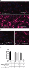
Figure 3. Specificity of Au-NPs for targeted, cancer cell specific mRNAs. A, B
Cy5-DNA Au-NPs targeting the human BIRC5 (A) or AML1/ETO (B) mRNAs were added to cells expressing doxycycline-inducible BIRC5 or AML1/ETO mRNA in the presence or absence of doxycycline. For these Au-NPs, the non-covalently linked oligonucleotide contained a Cy5 fluorophore rather than dasatinib. After 24 hours, the cells were stained with Hoechst 33342 dye and assessed by microscopy. Hoechst (blue) and Cy5 (red). Representative images are shown. C. Dasatinib-DNA Au-NPs targeting the human BIRC5 mRNA were added to recombinant SRC kinase in the presence of an excess of either scrambled oligonucleotide or an oligonucleotide mimicking the sequence of human BIRC5 mRNA. SRC kinase activity was measured using a luciferase-based peptide phosphorylation assay. Data are shown relative to the untreated control and are the mean±s.e.m. from three independent experiments. *, P < 0.0001 comparing the untreated control and dasatinib-DNA Au-NPs plus survivin oligonucleotide.
Figure 4
Figure 4. Highly efficient uptake of Au-NPs by leukemia cells
A. Percentage of leukemia cells containing DNA Au-NPs covalently labeled with Cy5 (see methods) was assessed by flow cytometry after an overnight incubation. Percentages are the mean±s.e.m. from three independent experiments. K562 cells were mixed with either human bone marrow mononuclear cells B. or murine bone marrow cells C. at the specified ratios and then incubated overnight with DNA Au-NPs covalently labeled with Cy5. Percentages of leukemia cells containing NPs are the mean±s.e.m. from three independent experiments. Flow cytometry histogram D. and quantitation E. of K562 leukemia cells isolated from subcutaneous flanks tumors that were either injected with Cy5 labeled Au-NPs (blue, green, orange) or un-injected (red).
Figure 5
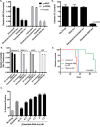
Figure 5. Dasatinib-DNA Au-NPs target human leukemia cells
A. K562 leukemia cells were treated with either dasatinib or dasatinib-DNA Au-NPs (1 nM) targeting either the human BIRC5 mRNA or a scrambled control sequence. After 24 hours the cells were fixed/permeabilized, stained for intracellular phospho-SRC or phospho-CRKL, and assessed by flow cytometry. Median fluorescence intensity (MFI) for each condition relative to the untreated control was calculated. *, P < 0.0001 for both phospho-SRC and phospho-CRKL when comparing untreated and either dasatinib or dasatinib-DNA Au-NP treated cells. B. Dasatinib-sensitive human leukemia cells (SKNO-1, Kasumi-1, and K562) and a dasatinib-insensitive murine hematopoietic cell line (32D) were treated with dasatinib-DNA Au-NPs and after 48 hours proliferation measured using the Cell-TiterGlo luciferase assay. The data are normalized to the untreated controls and are the mean±s.e.m. from three independent experiments. C. Induction of apoptosis (Annexin-V positive cells) in K562 leukemia cells 48 hours after treatment with either dasatinib (1 μM) or dasatinib-DNA Au-NPs at the specified concentrations. D. Colony formation of K562 leukemia cells treated for 4 hours with dasatinib-DNA Au-NPs, targeting either human BIRC5 mRNA or a scrambled control, or control NPs lacking dasatinib prior to plating in methylcellulose. Colonies were counted 7 days after seeding 103 cells/plate. Values are expressed as the percentage of colonies from drug treated cells compared to untreated control cells. *, P < 0.001 when comparing untreated control cells and cells treated with dasatinib-DNA Au-NPs. E. Survival curves for mice intravenously injected with K562 leukemia cells that were either untreated or treated for 4 hours prior to injection with dasatinib-DNA Au-NPs or control DNA-Au-NPs lacking dasatinib. N=3 for untreated and dasatinib-DNA Au-NP treated and N=2 for control NPs lacking dasatinib. *, P < 0.0001 when comparing the mice receiving dasatinib-DNA Au-NP treated cells versus untreated leukemia cells.
Figure 6
Figure 6. Dasatinib-DNA Au-NPs exhibit less toxicity than dasatinib alone against human CD34 and T-cells
A. Stimulated (SCF, FLT-3 ligand, thrombopoietin) human CD34 cells were treated with either dasatinib or dasatinib-DNA Au-NPs. After 24 hours, the cells were fixed/permeabilized, stained for intracellular phospho-SRC, and median fluorescent intensity (MFI) assessed by flow cytometry. The MFI values are normalized to the untreated controls and are the mean±s.e.m. from three independent experiments. **, P not significant. *, P < 0.001 comparing untreated and dasatinib treated cells. B. Human CD3+ T-cells were stimulated with anti-CD3/anti-CD28 beads in the absence of any drug or in the presence of either dasatinib or dasatinib-DNA Au-NPs. After 24 hours, T-cell activation was assessed by CD69 staining and flow cytometry. Data are the mean±s.e.m. from three independent experiments. *, P < 0.01 and **, P not significant when compared to untreated cells. C. Dasatinib-DNA Au-NPs targeting the human BIRC5 gene were added to murine NIH3T3 cells expressing doxycycline-inducible human BIRC5 mRNA in the presence or absence of doxycycline. After 24 hours, the cells were fixed/permeabilized, stained for intracellular phospho-SRC, and median fluorescent intensity (MFI) assessed by flow cytometry. The MFI values are the mean±s.e.m. from three independent experiments. *, P < 0.001 and **, P < 0.0001.

References

    1. Barnaby SN, Sita TL, Petrosko SH, Stegh AH, Mirkin CA. Therapeutic applications of spherical nucleic acids. Cancer Treat Res. 2015;166:23–50. doi: 10.1007/978-3-319-16555-4_2. - DOI - PubMed
    1. Rosi NL, Giljohann DA, Thaxton CS, Lytton-Jean AKR, Han MS, Mirkin CA. Oligonucleotide-modified gold nanoparticles for intracellular gene regulation. Science. 2006;312:1027–30. doi: 10.1126/science.1125559. - DOI - PubMed
    1. Prigodich AE, Seferos DS, Massich MD, Giljohann DA, Lane BC, Mirkin CA. Nano-flares for mRNA regulation and detection. ACS Nano. 2009;3:2147–52. doi: 10.1021/nn9003814. - DOI - PMC - PubMed
    1. Prigodich AE, Randeria PS, Briley WE, Kim NJ, Daniel WL, Giljohann DA, Mirkin CA. Multiplexed nanoflares: mRNA detection in live cells. Anal Chem. 2012;84:2062–6. doi: 10.1021/ac202648w. - DOI - PMC - PubMed
    1. Giljohann DA, Seferos DS, Daniel WL, Massich MD, Patel PC, Mirkin CA. Gold nanoparticles for biology and medicine. Angew Chem Int Ed Engl. 2010;49:3280–94. doi: 10.1002/anie.200904359. - DOI - PMC - PubMed

MeSH terms