. 2016 May 20;11(5):e0155033.
doi: 10.1371/journal.pone.0155033. eCollection 2016.

Characterisation of Candida within the Mycobiome/Microbiome of the Lower Respiratory Tract of ICU Patients

Affiliations

Characterisation of Candida within the Mycobiome/Microbiome of the Lower Respiratory Tract of ICU Patients

Robert Krause et al. PLoS One. .

Abstract

Whether the presence of Candida spp. in lower respiratory tract (LRT) secretions is a marker of underlying disease, intensive care unit (ICU) treatment and antibiotic therapy or contributes to poor clinical outcome is unclear. We investigated healthy controls, patients with proposed risk factors for Candida growth in LRT (antibiotic therapy, ICU treatment with and without antibiotic therapy), ICU patients with pneumonia and antibiotic therapy and candidemic patients (for comparison of truly invasive and colonizing Candida spp.). Fungal patterns were determined by conventional culture based microbiology combined with molecular approaches (next generation sequencing, multilocus sequence typing) for description of fungal and concommitant bacterial microbiota in LRT, and host and fungal biomarkes were investigated. Admission to and treatment on ICUs shifted LRT fungal microbiota to Candida spp. dominated fungal profiles but antibiotic therapy did not. Compared to controls, Candida was part of fungal microbiota in LRT of ICU patients without pneumonia with and without antibiotic therapy (63% and 50% of total fungal genera) and of ICU patients with pneumonia with antibiotic therapy (73%) (p<0.05). No case of invasive candidiasis originating from Candida in the LRT was detected. There was no common bacterial microbiota profile associated or dissociated with Candida spp. in LRT. Colonizing and invasive Candida strains (from candidemic patients) did not match to certain clades withdrawing the presence of a particular pathogenic and invasive clade. The presence of Candida spp. in the LRT rather reflected rapidly occurring LRT dysbiosis driven by ICU related factors than was associated with invasive candidiasis.


Conflict of interest statement

Competing Interests: The authors have declared that no competing interests exist.

Figures

Fig 1
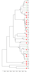
Fig 1. Neighbor-joining tree of invasive and colonizing Candida isolates.
Red dots = invasive Candida isolates; green circles = colonizing Candida isolates from patients without candidemia; red circles = oral colonizing isolates from patients with candidemia. The percentage of replicate trees in which the associated taxa clustered together in the bootstrap test (1000 replicates) are shown next to the branches. The tree is drawn to scale, with branch lengths in the same units as those of the evolutionary distances used to infer the phylogenetic tree. The evolutionary distances are in the units of the number of amino acid differences per site.
Fig 2
Fig 2. Relative abundance of fungal microbiota in lower respiratory tract samples.
Healthy adults (bar group 1a), patients with healthy respiratory tract but with antibiotic therapy for extrapulmonary infection (bar group 1b), non-neutropenic intubated and mechanically ventilated ICU patients without antibiotic therapy (bar group 2a), non-neutropenic intubated and mechanically ventilated ICU patients with antibiotic therapy for extrapulmonary infection (bar group 2b), and patients with antibiotic therapy due to pneumonia (bar group 3b). Fungi are shown at genus level. Yellow bars represent the sum of Candida sequences across all samples per group related to the total number of detected fungal sequences within each of the groups. By conventional culture Candida spp. were detected in 0/8 (group 1a), 0/7 (group 1b), 1/7 (group 2a), 3/6 (group 2b), and 11/26 (group 3b) patients. No other fungi were cultured.
Fig 3
Fig 3. Average relative Candida abundance in lower respiratory tract samples.
Healthy adults (bar group 1a), patients with healthy respiratory tract but with antibiotic therapy for extrapulmonary infection (bar group 1b), non-neutropenic intubated and mechanically ventilated ICU patients without antibiotic therapy (bar group 2a), non-neutropenic intubated and mechanically ventilated ICU patients with antibiotic therapy for extrapulmonary infection (bar group 2b), and patients with antibiotic therapy due to pneumonia (bar group 3b). Significant differences of average relative Candida abundance between groups are shown with bars representing the average Candida abundance and standard errors in given groups (* = p <0.05 for groups 2a, 2b, 3b versus 1a or 1b).
Fig 4
Fig 4. Relative abundance of bacterial microbiota in lower respiratory tract samples.
Healthy adults (bar group 1a), patients with healthy respiratory tract but with antibiotic therapy for extrapulmonary infection (bar group 1b), non-neutropenic intubated and mechanically ventilated ICU patients without antibiotic therapy (bar group 2a), non-neutropenic intubated and mechanically ventilated ICU patients with antibiotic therapy for extrapulmonary infection (bar group 2b), and patients with antibiotic therapy due to pneumonia (bar group 3b). Bacteria are shown at genus level. By conventional culture bacteria were cultured in 5/8 (group 1a), 2/7 (group 1b), 7/7 (group 2a), 3/6 (group 2b), and 13/26 (group 3b) patients.
Fig 5
Fig 5. Bray-Curtis distances between study groups indicating differences in fungal species composition.
Distance of zero indicates that groups are completely similar for every species. Distance of 1 indicates that groups are completely dissimilar and do not share any species. Group 1a, healthy adults; group 1b, healthy respiratory tract but with antibiotic therapy for extrapulmonary infection; group 2a, non-neutropenic intubated and mechanically ventilated ICU patients without antibiotic therapy; group 2b, non-neutropenic intubated and mechanically ventilated ICU patients with antibiotic therapy for extrapulmonary infection; group 3b, non-neutropenic intubated and mechanically ventilated ICU patients with antibiotic therapy due to pneumonia.
Fig 6
Fig 6. Bray-Curtis distances between study groups indicating differences in bacterial species composition.
Distance of zero indicates that groups are completely similar for every species. Distance of 1 indicates that groups are completely dissimilar and do not share any species. Group 1a, healthy adults; group 1b, healthy respiratory tract but with antibiotic therapy for extrapulmonary infection; group 2a, non-neutropenic intubated and mechanically ventilated ICU patients without antibiotic therapy; group 2b, non-neutropenic intubated and mechanically ventilated ICU patients with antibiotic therapy for extrapulmonary infection; group 3b, non-neutropenic intubated and mechanically ventilated ICU patients with antibiotic therapy due to pneumonia.
Fig 7
Fig 7. Associations/dissociations between bacteria and fungi (heatmaps).
Relationships (association/dissociation) between bacteria and fungi in lower respiratory tract samples of non-neutropenic intubated and mechanically ventilated ICU patients without antibiotic therapy calculated as odds ratios and depicted as heatmap. An odds ratio above 2 was considered a positive association (in red), an odds ratio below 0.5 was interpreted as negative association (= dissociation, in green). The scale shows odds ratios.
Fig 8
Fig 8. Associations/dissociations between bacteria and fungi (heatmaps).
Relationships (association/dissociation) between bacteria and fungi in lower respiratory tract samples of non-neutropenic intubated and mechanically ventilated ICU patients with antibiotic therapy for extrapulmonary infection calculated as odds ratios and depicted as heatmap. An odds ratio above 2 was considered a positive association (in red), an odds ratio below 0.5 was interpreted as negative association (= dissociation, in green). The scale shows odds ratios.
Fig 9
Fig 9. Associations/dissociations between bacteria and fungi (heatmaps).
Relationships (association/dissociation) between bacteria and fungi in lower respiratory tract samples of non-neutropenic intubated and mechanically ventilated ICU patients with antibiotic therapy due to pneumonia calculated as odds ratios and depicted as heatmap. An odds ratio above 2 was considered a positive association (in red), an odds ratio below 0.5 was interpreted as negative association (= dissociation, in green). The scale shows odds ratios. Asterisks show significant differences (p<0.05; 95% confidence interval) for odds ratios (Fig 9).
Fig 10
Fig 10. Relative abundance of fungal microbiota in consecutively sampled patients.
Consecutive lower respiratory tract samples of 4 intubated and mechanically ventilated patients with pneumonia. Fungi are shown at genus level. Yellow bars represent Candida species. VAP = Ventilator associated pneumonia, ASP = aspiration pneumonia, NAP = nosocomial acquired pneumonia.
Fig 11
Fig 11. Relative abundance of bacterial microbiota in consecutively sampled patients.
Consecutive lower respiratory tract samples of 4 intubated and mechanically ventilated patients with pneumonia. Bacteria are shown at genus level. Unclassified sequences in sample 304-3B-ASP-0 revealed Escherichia/Shigella sp. using a confidence threshold of 0.63. Comparison of these sequences with reference sequences using BLAST yielded uncultured Shigella sp. 16s rRNA genes. Unclassified sequences in sample 313-3B-VAP-0 revealed Enterobacter sp. using a confidence threshold of 0.63. Comparison of these sequences with reference sequences using BLAST yielded uncultured proteobacterium 16s rRNA genes. VAP = Ventilator associated pneumonia, ASP = aspiration pneumonia, NAP = nosocomial acquired pneumonia.

References

    1. Baum GL. The Significance of Candida albicans in Human Sputum. The Significance of Candida albicans in Human Sputum. New Engl J Med 1960; 263:70–3. - PubMed
    1. el-Ebiary M, Torres A, Fàbregas N, la Bellacasa de JP, González J, Ramirez J, et al. Significance of the isolation of Candida species from respiratory samples in critically ill, non-neutropenic patients. An immediate postmortem histologic study. Am J Respir Crit Care Med. 1997;156: 583–590. 10.1164/ajrccm.156.2.9612023 - DOI - PubMed
    1. Meersseman W, Lagrou K, Spriet I, Maertens J, Verbeken E, Peetermans WE, et al. Significance of the isolation of Candida species from airway samples in critically ill patients: a prospective, autopsy study. Intensive Care Med. 2009;35: 1526–1531. 10.1007/s00134-009-1482-8 - DOI - PubMed
    1. Rello J, Esandi ME, Díaz E, Mariscal D, Gallego M, Vallès J. The role of Candida sp isolated from bronchoscopic samples in nonneutropenic patients. Chest. 1998;114: 146–149. - PubMed
    1. Delisle M-S, Williamson DR, Perreault MM, Albert M, Jiang X, Heyland DK. The clinical significance of Candida colonization of respiratory tract secretions in critically ill patients. Journal of Critical Care. 2008;23: 11–17. 10.1016/j.jcrc.2008.01.005 - DOI - PubMed

MeSH terms

Substances

Supplementary concepts