Differential expression of pro-inflammatory and oxidative stress mediators induced by nitrogen dioxide and ozone in primary human bronchial epithelial cells
- PMID: 27206323
- PMCID: PMC4967931
- DOI: 10.1080/08958378.2016.1185199
Differential expression of pro-inflammatory and oxidative stress mediators induced by nitrogen dioxide and ozone in primary human bronchial epithelial cells
Abstract
Context: NO2 and O3 are ubiquitous air toxicants capable of inducing lung damage to the respiratory epithelium. Due to their oxidizing capabilities, these pollutants have been proposed to target specific biological pathways, but few publications have compared the pathways activated.
Objective: This work will test the premise that NO2 and O3 induce toxicity by activating similar cellular pathways.
Methods: Primary human bronchial epithelial cells (HBECs, n = 3 donors) were exposed for 2 h at an air-liquid interface to 3 ppm NO2, 0.75 ppm O3, or filtered air and harvested 1 h post-exposure. To give an overview of pathways that may be influenced by each exposure, gene expression was measured using PCR arrays for toxicity and oxidative stress. Based on the results, genes were selected to quantify whether expression changes were changed in a dose- and time-response manner using NO2 (1, 2, 3, or 5 ppm), O3 (0.25, 0.50, 0.75, or 1.00 ppm), or filtered air and harvesting 0, 1, 4 and 24 h post-exposure.
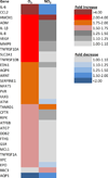
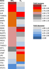
Results: Using the arrays, genes related to oxidative stress were highly induced with NO2 while expression of pro-inflammatory and vascular function genes was found subsequent to O3. NO2 elicited the greatest HMOX1 response, whereas O3 more greatly induced IL-6, IL-8 and PTGS2 expression. Additionally, O3 elicited a greater response 1 h post-exposure and NO2 produced a maximal response after 4 h.
Conclusion: We have demonstrated that these two oxidant gases stimulate differing mechanistic responses in vitro and these responses occur at dissimilar times.
Keywords: In vitro; inflammation; nitrogen dioxide; oxidant; oxidative stress; ozone.
Figures


Similar articles
-
Effect of ozone and nitrogen dioxide on the release of proinflammatory mediators from bronchial epithelial cells of nonatopic nonasthmatic subjects and atopic asthmatic patients in vitro.J Allergy Clin Immunol. 2001 Feb;107(2):287-94. doi: 10.1067/mai.2001.111141. J Allergy Clin Immunol. 2001. PMID: 11174195
-
Effect of ozone and nitrogen dioxide on the permeability of bronchial epithelial cell cultures of non-asthmatic and asthmatic subjects.Clin Exp Allergy. 2002 Sep;32(9):1285-92. doi: 10.1046/j.1365-2745.2002.01435.x. Clin Exp Allergy. 2002. PMID: 12220465
-
Pulmonary arachidonic acid metabolism following acute exposures to ozone and nitrogen dioxide.J Toxicol Environ Health. 1990 Dec;31(4):275-90. doi: 10.1080/15287399009531456. J Toxicol Environ Health. 1990. PMID: 2147723
-
Critical review of the human data on short-term nitrogen dioxide (NO2) exposures: evidence for NO2 no-effect levels.Crit Rev Toxicol. 2009;39(9):743-81. doi: 10.3109/10408440903294945. Crit Rev Toxicol. 2009. PMID: 19852560 Review.
-
The toxicity of air pollution in experimental animals and humans: the role of oxidative stress.Toxicol Lett. 1994 Jun;72(1-3):269-77. doi: 10.1016/0378-4274(94)90038-8. Toxicol Lett. 1994. PMID: 8202941 Review.
Cited by
-
Satellite-detected tropospheric nitrogen dioxide and spread of SARS-CoV-2 infection in Northern Italy.Sci Total Environ. 2020 Oct 15;739:140278. doi: 10.1016/j.scitotenv.2020.140278. Epub 2020 Jun 16. Sci Total Environ. 2020. PMID: 32758963 Free PMC article.
-
Association of coronavirus pathogencity with the level of antioxidants and immune system.J Family Med Prim Care. 2021 Feb;10(2):609-614. doi: 10.4103/jfmpc.jfmpc_1007_20. Epub 2021 Feb 27. J Family Med Prim Care. 2021. PMID: 34041049 Free PMC article. Review.
-
Transcriptional Effects of Ozone and Impact on Airway Inflammation.Front Immunol. 2019 Jul 10;10:1610. doi: 10.3389/fimmu.2019.01610. eCollection 2019. Front Immunol. 2019. PMID: 31354743 Free PMC article. Review.
-
Long-term air pollution exposure and incident stroke in American older adults: A national cohort study.Glob Epidemiol. 2022 Dec;4:100073. doi: 10.1016/j.gloepi.2022.100073. Epub 2022 Apr 16. Glob Epidemiol. 2022. PMID: 36644436 Free PMC article.
-
Beyond allergic progression: From molecules to microbes as barrier modulators in the gut-lung axis functionality.Front Allergy. 2023 Jan 30;4:1093800. doi: 10.3389/falgy.2023.1093800. eCollection 2023. Front Allergy. 2023. PMID: 36793545 Free PMC article. Review.
References
-
- Ayyagari VN, Januszkiewicz A, Nath J. Pro-inflammatory responses of human bronchial epithelial cells to acute nitrogen dioxide exposure. Toxicology. 2004;197:149–164. - PubMed
-
- Ayyagari VN, Januszkiewicz A, Nath J. Effects of nitrogen dioxide on the expression of intercellular adhesion molecule-1, neutrophil adhesion, and cytotoxicity: studies in human bronchial epithelial cells. Inhal Toxicol. 2007;19:181–194. - PubMed
-
- Ballinger CA, Cueto R, Squadrito G, Coffin JF, Velsor LW, Pryor WA, Postlethwait EM. Antioxidant-mediated augmentation of ozone-induced membrane oxidation. Free Radic Biol Med. 2005;38:515–526. - PubMed
Publication types
MeSH terms
Substances
Grants and funding
LinkOut - more resources
Full Text Sources
Other Literature Sources
Medical
Research Materials
