Review
. 2017 Jan 1;81(1):31-42.
doi: 10.1016/j.biopsych.2016.03.2104. Epub 2016 Mar 31.

Pathway-Specific Dopamine Abnormalities in Schizophrenia

Affiliations
Review

Pathway-Specific Dopamine Abnormalities in Schizophrenia

Jodi J Weinstein et al. Biol Psychiatry. .

Abstract

In light of the clinical evidence implicating dopamine in schizophrenia and the prominent hypotheses put forth regarding alterations in dopaminergic transmission in this disease, molecular imaging has been used to examine multiple aspects of the dopaminergic system. We review the imaging methods used and compare the findings across the different molecular targets. Findings have converged to suggest early dysregulation in the striatum, especially in the rostral caudate, manifesting as excess synthesis and release. Recent data showed deficit extending to most cortical regions and even to other extrastriatal subcortical regions not previously considered to be "hypodopaminergic" in schizophrenia. These findings yield a new topography for the dopaminergic dysregulation in schizophrenia. We discuss the dopaminergic innervation within the individual projection fields to provide a topographical map of this dual dysregulation and explore potential cellular and circuit-based mechanisms for brain region-dependent alterations in dopaminergic parameters. This refined knowledge is essential to better guide translational studies and efforts in early drug development.

Keywords: Cortex; Dopamine; Neuroanatomy; PET imaging; Schizophrenia; Striatum.


Conflict of interest statement

Dr. Kegeles has received research support from Amgen. Dr. Slifstein has received research support from Forest Laboratories, Pierre-Fabre, CHDI, and Otsuka and has provided consultation for Amgen. Dr. Abi-Dargham has received research support from Takeda and Forest Pharmaceuticals and has served on advisory boards for Roche, Forum, and Otsuka. Drs. Moore, Chohan, and Weinstein report no biomedical financial interests or potential conflicts of interest.

Figures

Figure 1
Figure 1. Dopaminergic Imaging Targets
Schematic of imaging methods used to measure aspects of the dopamine (DA) system in vivo. Graphic depicts progression of DA from synthesis (A), storage (B), to release (E,F), then either reuptake by dopamine transporter (DAT, C) or binding to receptor (D). Imaging targets and related paradigms are described in accompanying text.
Figure 2
Figure 2. Topography of dopaminergic innervation and receptor distribution
Schematic representation of distributions of dopamine D1 and D2 receptors (left hemispheres) and patterns of dopaminergic innervations (right hemispheres) in select primate (left panel) and rodent (right panel) brain regions. Left hemispheres: Brown and black squares depict D1 and D2 receptors, respectively. Throughout the primate and rodent brain, D1 receptors (D1) are present at a higher density than D2 receptors (D2). The striatum, and in particular the caudate-putamen, has the highest densities of dopamine (DA) receptors. DA receptors are also present in medium-to-low densities in the cortex, pallidum and midbrain. Receptor densities are relatively low in thalamus, amygdala and hippocampus. See text for details. Right hemispheres: Topographical distribution of DA cell bodies (filled circles) and their terminals (lines). In the primate panel, red circles represent DA cell bodies in the VTA with terminals in the cortex, striatum (in particular the ventral part), pallidum, thalamus and amygdala. The VTA dopaminergic cellular organization is better characterized in the rodent where discrete VTA cell groups project to the cortex (red), nucleus accumbens (dark green) and amygdala (orange). In the primate, SN dorsal tier cell group (light green) projects to the cortex and ventral striatum, as well as the pallidum, thalamus and amygdala. The rodent brain in contrast has a low density of these dorsal tier neurons. The SN ventral tier groups (SN compacta densocellular part (dark blue) and fingers (light blue)) project heavily and topographically to caudate-putamen with medium/low innervations of cortex, ventral striatum, thalamus and amygdala. See text for further details.
Figure 3
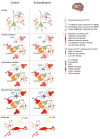
Figure 3. Topography of dopamine release findings in schizophrenia compared to controls
Schematic representations of DA release characteristics in the cortex (top), striatum (middle) and midbrain (bottom) in healthy controls (HC) and patients with schizophrenia (SZ) based on imaging findings in patients. DA neuron cell bodies, terminals and transmitters are depicted in red. Color gradients depict DA terminal densities. Cortex: The cortex receives sparse dopaminergic innervation that is poor in dopamine D2 receptors (D2) and transporter expression. This sculpts D2 displacement measurement, which is low in the cortex. In schizophrenia there is evidence for reduced cortical DA release. See text for details. Striatum: DA and cortical neuron terminals (green) are shown innervating medium spiny neuron spines (orange). Also shown are local cholinergic (blue) and GABAergic (brown) interneuron populations forming the striatal microcircuitry. There is considerable heterogeneity in DA release across striatal regions, e.g. dopaminergic innervation of ventral striatum (VST, also referred to as LST) is relatively sparse and is derived from dorsal tier cell groups that are poor in D2 and DAT. In contrast the sensorimotor striatum (SMST) receives dense dopaminergic inputs mostly from the ventral tier DA neurons that are rich in D2 and DAT. A greater number of synapse sites in the ventral striatum and high levels of D2 and DAT in SMST may account for high D2 displacement in these regions. Compared to VST and SMST, stimulant induced D2 displacement is low in the associative striatum (AST). In schizophrenia, DA release is increased across substriatal divisions due to a prominent increase in the AST. Midbrain: Shown are DA cell bodies, local GABAergic interneurons (brown) and D1 medium spiny neuron terminals (yellow). While there is heterogeneity in the level of expression of D2 receptors and DAT (e.g. dorsal tier and especially medial VTA neurons have low D2 and DAT levels), imaging studies showing subregional analysis of D2 displacement are lacking. However, in SZ there is a reduced stimulant-induced D2 displacement.
Figure 4
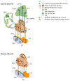
Figure 4. Striatal patch-matrix connectome
Schematic representation of striatal patch-matrix connectome. Afferents: The cortex topographically projects to the striatum. Within the cortex deeper cortical layers innervate striatal patches (dark brown) whereas the surrounding matrix (light brown) is innervated by superficial cortical layers (light brown). Within the midbrain, the dorsal tier (orange and yellow) innervates the matrix, as do the non-dopaminergic cells (dark green) from the same region. Patch innervation from the midbrain is mostly derived from the ventral tier cell groups (dark blue). Non-dopaminergic (presumably GABAergic) projection neurons within the SNr innervate the striatal matrix complex. Efferents: Striatal patch neurons (maroon) mostly project to ventral tier DA cells. These include both D1 receptor expressing medium spiny neurons and other striatal projection neurons. Striatal projection neurons within the matrix project to both DA and non-dopaminergic populations within the dorsal tier and GABAergic populations in the SNr. See text for further details.

References

    1. Rossum V. The significance of dopamine receptor blockade for the mechanism of action of neuroleptic drugs. Arch Int Pharmacodyn Therapy. 1966;160:492–494. - PubMed
    1. Carlsson A. Antipsychotic drugs, neurotransmitters, and schizophrenia. The American journal of psychiatry. 1978;135:165–173. - PubMed
    1. Seeman P, Lee T, Chau-Wong M, Wong K. Antipsychotic drug doses and neuroleptic/dopamine receptors. Nature. 1976;261:717–719. - PubMed
    1. Lieberman JA, Kane JM, Alvir J. Provocative tests with psychostimulant drugs in schizophrenia. Psychopharmacology. 1987;91:415–433. - PubMed
    1. Weinberger DR, Berman KF. Speculation on the meaning of cerebral metabolic hypofrontality in schizophrenia. Schizophrenia Bull. 1988;14:157–168. - PubMed

Publication types

MeSH terms

Substances