. 2016 Jul;65(7):2006-19.
doi: 10.2337/db15-0340. Epub 2016 Apr 8.

Mast Cells Regulate Wound Healing in Diabetes

Affiliations

Mast Cells Regulate Wound Healing in Diabetes

Ana Tellechea et al. Diabetes. 2016 Jul.

Abstract

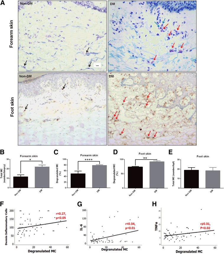
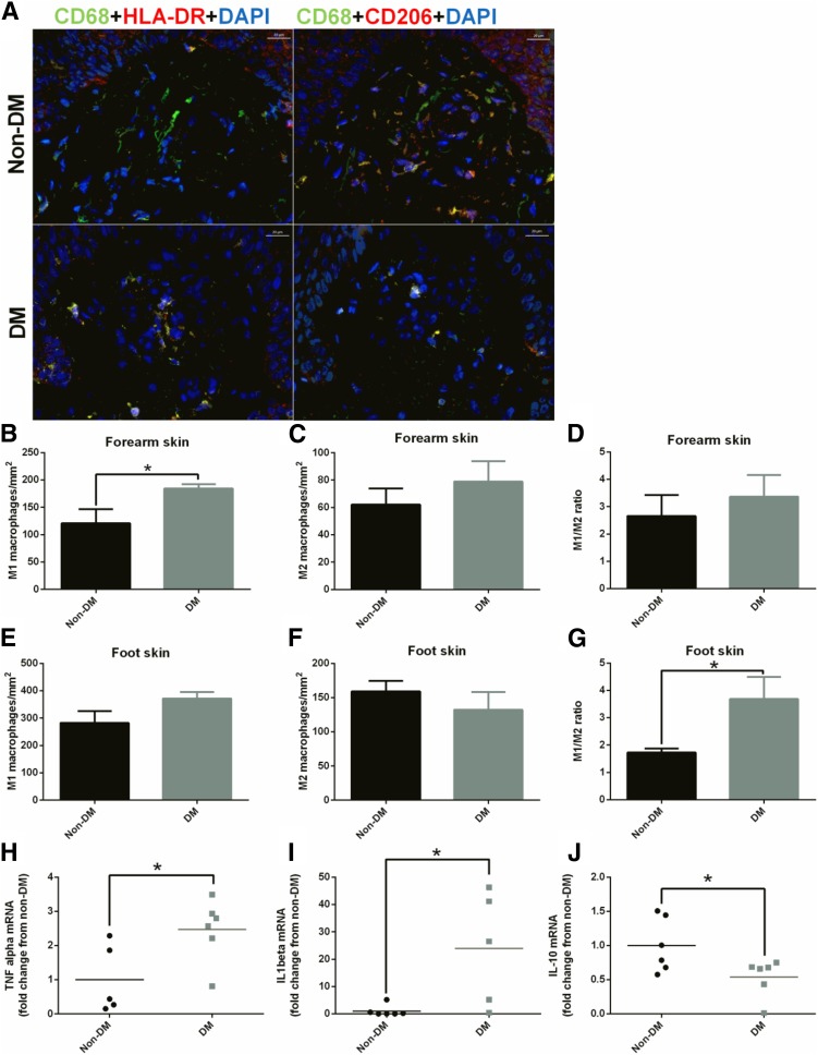
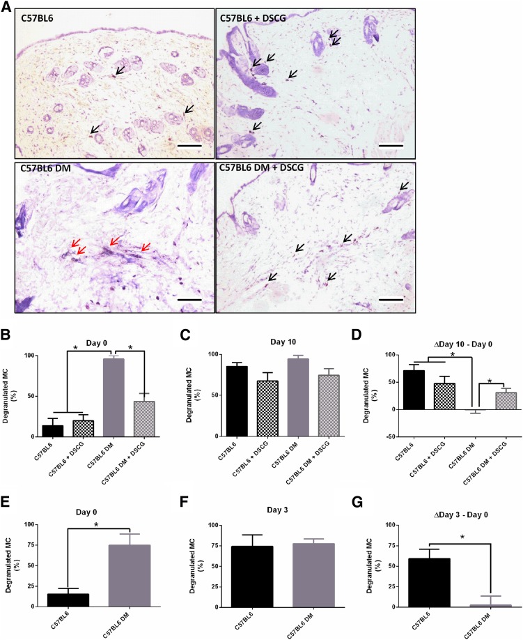
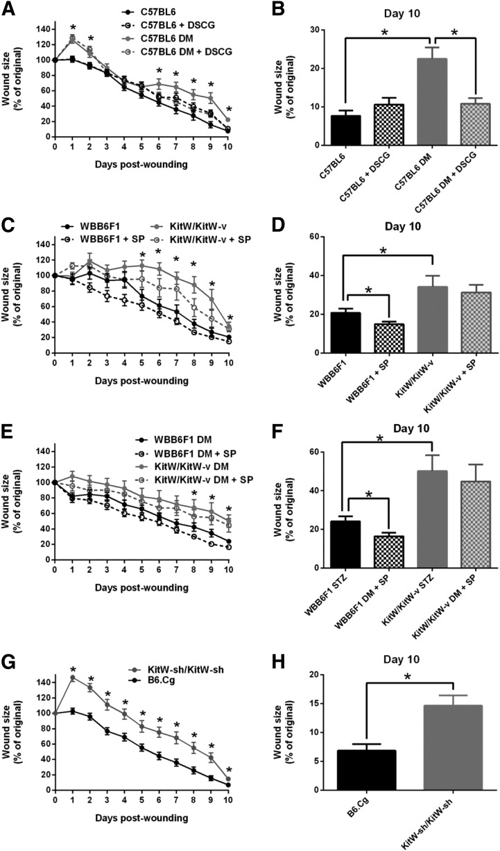
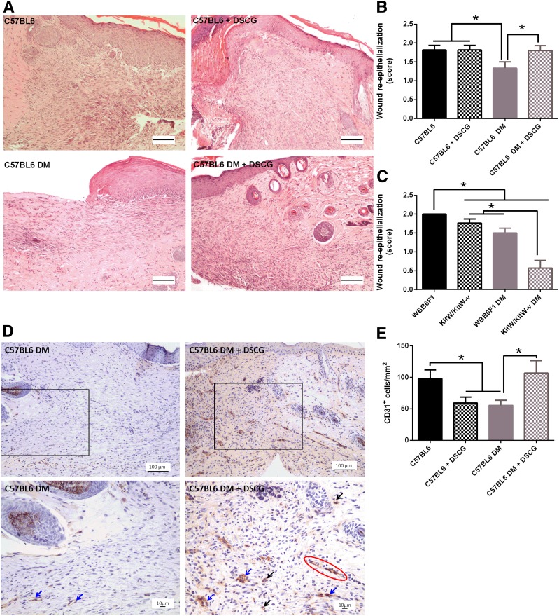
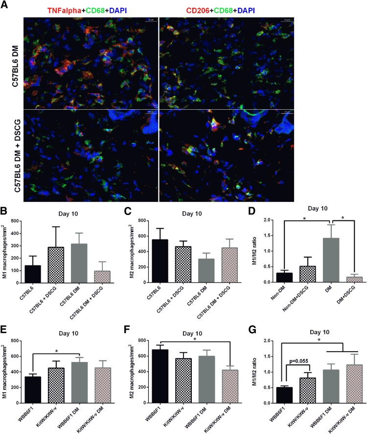
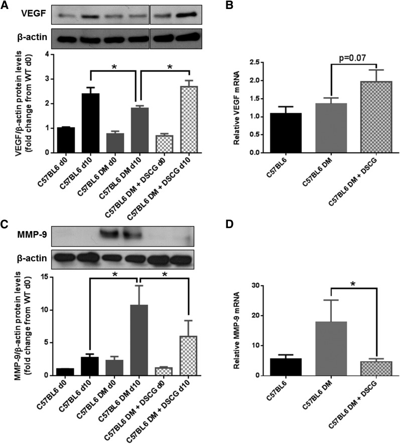
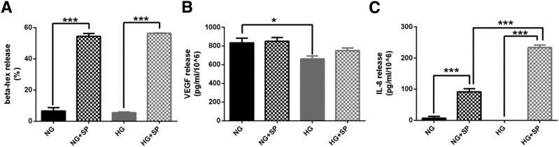
Diabetic foot ulceration is a severe complication of diabetes that lacks effective treatment. Mast cells (MCs) contribute to wound healing, but their role in diabetes skin complications is poorly understood. Here we show that the number of degranulated MCs is increased in unwounded forearm and foot skin of patients with diabetes and in unwounded dorsal skin of diabetic mice (P < 0.05). Conversely, postwounding MC degranulation increases in nondiabetic mice, but not in diabetic mice. Pretreatment with the MC degranulation inhibitor disodium cromoglycate rescues diabetes-associated wound-healing impairment in mice and shifts macrophages to the regenerative M2 phenotype (P < 0.05). Nevertheless, nondiabetic and diabetic mice deficient in MCs have delayed wound healing compared with their wild-type (WT) controls, implying that some MC mediator is needed for proper healing. MCs are a major source of vascular endothelial growth factor (VEGF) in mouse skin, but the level of VEGF is reduced in diabetic mouse skin, and its release from human MCs is reduced in hyperglycemic conditions. Topical treatment with the MC trigger substance P does not affect wound healing in MC-deficient mice, but improves it in WT mice. In conclusion, the presence of nondegranulated MCs in unwounded skin is required for proper wound healing, and therapies inhibiting MC degranulation could improve wound healing in diabetes.


Figures

Figure 1
Figure 1
Skin MC degranulation, assessed by toluidine blue and/or tryptase immunostaining, is increased in patients with diabetes and is associated with inflammation. A: Representative images of toluidine blue–stained MCs in forearm skin (top panel) and of tryptase-immunostained MCs in foot skin specimens (bottom panel) from subjects without diabetes (Non-DM) and with diabetes (DM). Scale bar: 10 µm. Black arrows show nondegranulated MCs, and red arrows show degranulated MCs. Degranulated MCs were in proximity with inflammatory cells (blue arrows). The total number (B) and percentage (C) of degranulated MCs stained with toluidine blue were increased in forearm skin specimens from patients with diabetes. MC degranulation was also increased in foot skin specimens from subjects with diabetes stained with tryptase (D), while the total number of MCs was not different (E). *P < 0.05, **P < 0.01, and ****P < 0.0001. A positive correlation was observed between degranulated MCs and the dermis inflammatory cells (F), and the serum levels of IL-6 (G) and TNF-α (H). hpf, high power field (400× magnification).
Figure 2
Figure 2
Skin macrophage phenotype is polarized toward M1 in patients with diabetes. A: Representative images of M1 and M2 macrophages in foot skin specimens from subjects without diabetes (Non-DM) and with diabetes (DM). Scale bar: 20 µm. M1 (left panel CD68+HLADR+DAPI) and M2 (right panel CD68+CD206+DAPI) are shown in yellow-orange as a result from the triple-positive staining. In the forearm skin, the number of M1 macrophages was increased in subjects with diabetes (B), whereas the number of M2 macrophages (C) and the M1/M2 ratio (D) were not different. In the foot skin, the number of M1 macrophages tended to be increased in subjects with diabetes (E), whereas the number of M2 macrophages tended to be reduced (F), resulting in a higher M1/M2 ratio (G). mRNA expression of the M1 markers TNF-α (H) and IL-1β (I) was increased, whereas the expression of M2 marker IL-10 was reduced (J) in the foot skin of patients with diabetes. *P < 0.05.
Figure 3
Figure 3
MC degranulation is increased in unwounded skin of diabetic (DM) mice and fails to further increase after wounding. A: Representative images of nondegranulated (black arrows) and degranulated (red arrows) MCs in day 0 skin biopsy specimens from C57BL/6J nondiabetic and diabetic mice, untreated and pretreated with DSCG. Scale bar: 100 μm. B: MC degranulation was increased in diabetic mice at day 0 and after intraperitoneal DSCG treatment for 10 days before wounding reduced it. C: Nondiabetic mice showed increased MC degranulation at day 10 when compared with day 0, but there were no changes in diabetic mice between days 0 and 10. As a result, there were no differences among the various groups at day 10. D: When the difference between days 10 and 0 was calculated, a significant increase was noticed in nondiabetic mice, regardless of whether they were treated with DSCG or not. However, no difference was observed in the diabetic mice not treated with DSCG, but this difference was restored in the DSCG-treated diabetic mice. E–G: Similar results were observed in a different set of nondiabetic and diabetic mice that were studied at day 3 postwounding. Thus, MC degranulation was also increased in the skin in diabetic mice on day 0 (E), but was not different at day 3 (F), resulting in a failure to increase MC degranulation from day 0 to day 3 postwounding in diabetic mice (G). *P < 0.05.
Figure 4
Figure 4
Functional MCs are required for proper wound healing. Wound-healing progress was evaluated over a 10-day period in nondiabetic and diabetic mice treated with DSCG or not treated and MC-deficient mice. A and B: Wound healing was delayed in C57BL6 diabetic mice compared with nondiabetic mice and DSCG pretreatment accelerated it from days 6 to 10 postwounding. DSCG had no effect on nondiabetic mice. C–F: Wound healing was delayed in KitW/KitW-v mice without or with diabetes, when compared with their respective nondiabetic or diabetic WBB6F1 controls. Topical SP improved healing at day 10 postwounding in both nondiabetic and diabetic WBB6F1 mice, but failed to have an effect in either nondiabetic KitW/KitW-v or diabetic KitW/KitW-v mice. G and H: Wound healing was delayed in MC-deficient KitW-sh/KitW-sh mice when compared with their respective B6.Cg controls. *P < 0.05. DM, diabetic.
Figure 5
Figure 5
Wound re-epithelialization and neovascularization are reduced in diabetic (DM) mice and in KitW/KitW-v mice, and DSCG improves them in diabetic mice. Wound re-epithelialization and neovascularization were evaluated by histomorphometric analysis and CD31 staining, respectively. A: Representative hematoxylin-eosin images of C57BL6 mice, C57BL6 mice pretreated with DSCG, C57BL6 diabetic mice, and C57BL6 diabetic mice pretreated with DSCG. Scale bar: 100 µm. B: Wound re-epithelialization was reduced in C57BL6 diabetic mice when compared with both nondiabetic and diabetic DSCG-treated mice. C: Wound re-epithelialization was reduced in WBB6F1 diabetic mice when compared with their nondiabetic controls. Nondiabetic and diabetic KitW/KitW-v mice had lower re-epithelialization than their respective nondiabetic and diabetic WBB6F1 controls. Diabetic KitW/KitW-v mouse wounds had more incomplete re-epithelialization than nondiabetic KitW/KitW-v wounds. D: Representative images using CD31 staining of untreated C57BL6 diabetic mice and C57BL6 diabetic mice pretreated with DSCG, at lower (top panel) and higher (bottom panel) magnification. The red circle shows a vascular sprout, blue arrows show small-caliber blood vessels with an open lumen, and black arrows show either single CD31+ cells or groups of two to three CD31+ cells. E: Single CD31+ cells were reduced in nondiabetic DSCG-treated mice and C57BL6 diabetic mice compared with C57BL6 nondiabetic mice. Diabetic DSCG-treated mice had similar numbers of CD31+ cells compared with nondiabetic controls. *P < 0.05.
Figure 6
Figure 6
M1/M2 macrophage ratio is increased in the periwound skin of diabetic mice, and DSCG pretreatment restores it to normal levels. A: Representative image of M1 and M2 macrophages from day 10 periwound skin of C57BL6 diabetic mice, nontreated mice, and DSCG pretreated mice. Scale bar: 20 μm. M1 (left panel, CD68+TNF-α+DAPI) and M2 (right panel, CD68+CD206+DAPI) are shown in yellow-orange as a result from the triple-positive staining. B: C57BL6 diabetic mice tended to have elevated numbers of M1 macrophages compared with both C57BL6 nondiabetic (Non-DM) and diabetic (DM) mice pretreated with DSCG. C: C57BL6 diabetic mice tended to have reduced numbers of M2 macrophages compared with both C57BL6 nondiabetic and diabetic mice pretreated with DSCG. D: C57BL6 diabetic mice had a significantly higher periwound M1/M2 ratio than their respective nondiabetic controls. DSCG pretreatment restored the M1/M2 ratio to normal levels in diabetic mice but had no effect in nondiabetic mice. E: Diabetic WBB6F1 mice had elevated numbers of M1 macrophages compared with their nondiabetic controls. Nondiabetic and diabetic KitW/KitW-v mice tended to have higher counts of M1 macrophages than nondiabetic WBB6F1 mice. F: Diabetic KitW/KitW-v mice had reduced numbers of M2 macrophages. Nondiabetic KitW/KitW-v and diabetic WBB6F1 mice tended to have reduced numbers of M2 macrophages when compared with nondiabetic WBB6F1 mice. G: Diabetic WBB6F1 and diabetic KitW/KitW-v mice had higher M1/M2 ratios in the periwound skin compared with nondiabetic WBB6F1 mice. Nondiabetic KitW/KitW-v mice had a marginally increased M1/M2 ratio compared with WBB6F1 mice (P = 0.55). *P < 0.05.
Figure 7
Figure 7
DSCG pretreatment increases VEGF and reduces MMP-9 skin expression in diabetic mice. Day 0 and day 10 VEGF and MMP-9 skin protein and mRNA expression from C57BL6 nondiabetic mice, nontreated C57BL6 diabetic mice (DM), and DSCG-treated C57BL6 diabetic mice were evaluated by Western blot and qRT-PCR, respectively. A: VEGF protein expression was reduced in wounds in diabetic C57BL6 mice on day 10, but DSCG pretreatment restored it to levels observed in nondiabetic mice. B: DSCG pretreatment marginally increased VEGF mRNA in C57BL6 diabetic mice (P = 0.07). C: MMP-9 protein expression was increased in both day 0 skin and day 10 wounds from C57BL6 diabetic mice when compared with their C57BL6 nondiabetic controls, and DSCG pretreatment reduced it. D: DSCG pretreatment reduced MMP-9 mRNA in C57BL6 diabetic mice. *P < 0.05 (comparisons shown in graph).
Figure 8
Figure 8
HG does not affect MC degranulation but influences the release of MC mediators. A: SP increased β-hex release from LAD2 cells cultured in NG or HG conditions. HG level did not have any effect on β-hex release. B: HG level reduced VEGF release from LAD2 cells. SP did not affect VEGF release from LAD2 cells, either in NG or HG conditions. C: SP increased IL-8 release from LAD2 cells cultured in either NG or HG conditions. No differences were observed between NG and HG conditions alone. However, IL-8 release from LAD2 cells in the HG condition and triggered with SP was 61% higher than that in the low-glucose plus SP condition. *P < 0.05, ***P < 0.001.

References

    1. Diabetes: 1996 Vital Statistics. Vol 31 Alexandria, VA, American Diabetes Association, 1996
    1. Ramsey SD, Newton K, Blough D, et al. . Incidence, outcomes, and cost of foot ulcers in patients with diabetes. Diabetes Care 1999;22:382–387 - PubMed
    1. Falanga V. Wound healing and its impairment in the diabetic foot. Lancet 2005;366:1736–1743 - PubMed
    1. Singer AJ, Clark RA. Cutaneous wound healing. N Engl J Med 1999;341:738–746 - PubMed
    1. Galkowska H, Olszewski WL, Wojewodzka U, Rosinski G, Karnafel W. Neurogenic factors in the impaired healing of diabetic foot ulcers. J Surg Res 2006;134:252–258 - PubMed

Publication types