. 2016 Jun;65(6):1590-600.
doi: 10.2337/db15-1434. Epub 2016 Apr 5.

Integrin-Linked Kinase in Muscle Is Necessary for the Development of Insulin Resistance in Diet-Induced Obese Mice

Affiliations

Integrin-Linked Kinase in Muscle Is Necessary for the Development of Insulin Resistance in Diet-Induced Obese Mice

Li Kang et al. Diabetes. 2016 Jun.

Abstract

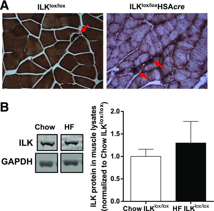
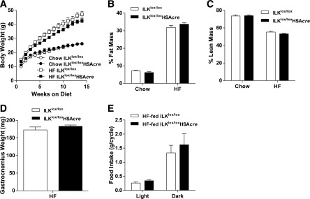
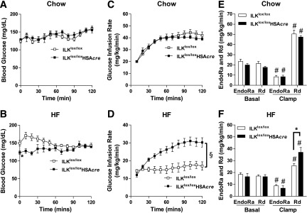
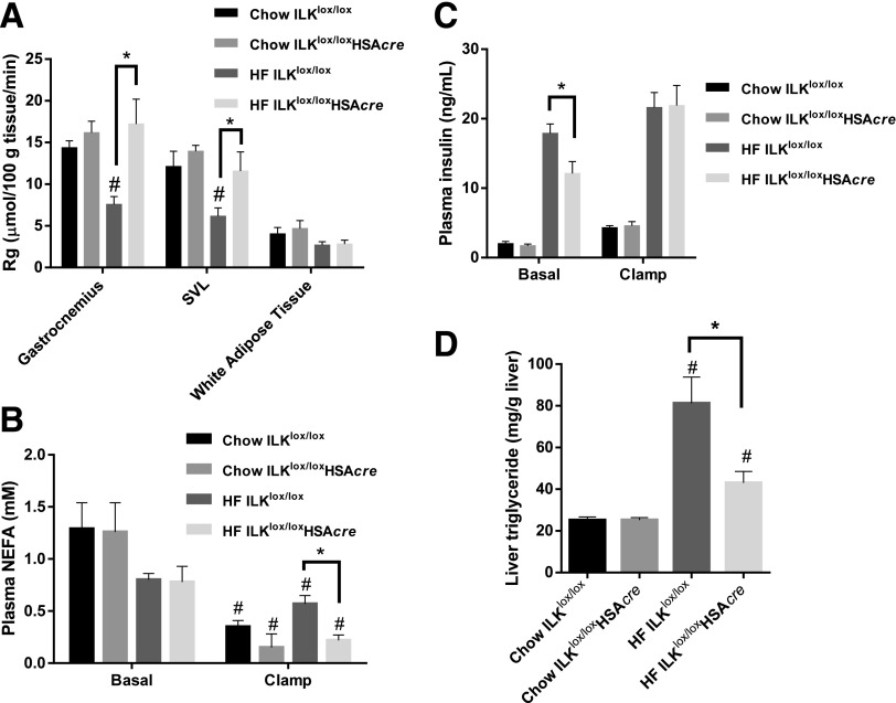
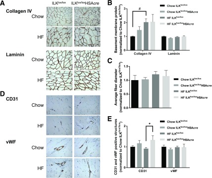
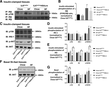
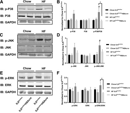
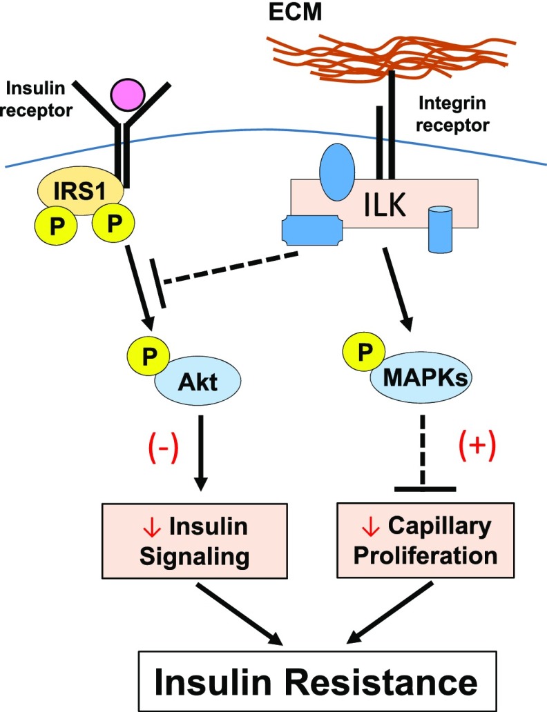
Diet-induced muscle insulin resistance is associated with expansion of extracellular matrix (ECM) components, such as collagens, and the expression of collagen-binding integrin, α2β1. Integrins transduce signals from ECM via their cytoplasmic domains, which bind to intracellular integrin-binding proteins. The integrin-linked kinase (ILK)-PINCH-parvin (IPP) complex interacts with the cytoplasmic domain of β-integrin subunits and is critical for integrin signaling. In this study we defined the role of ILK, a key component of the IPP complex, in diet-induced muscle insulin resistance. Wild-type (ILK(lox/lox)) and muscle-specific ILK-deficient (ILK(lox/lox)HSAcre) mice were fed chow or a high-fat (HF) diet for 16 weeks. Body weight was not different between ILK(lox/lox) and ILK(lox/lox)HSAcre mice. However, HF-fed ILK(lox/lox)HSAcre mice had improved muscle insulin sensitivity relative to HF-fed ILK(lox/lox) mice, as shown by increased rates of glucose infusion, glucose disappearance, and muscle glucose uptake during a hyperinsulinemic-euglycemic clamp. Improved muscle insulin action in the HF-fed ILK(lox/lox)HSAcre mice was associated with increased insulin-stimulated phosphorylation of Akt and increased muscle capillarization. These results suggest that ILK expression in muscle is a critical component of diet-induced insulin resistance, which possibly acts by impairing insulin signaling and insulin perfusion through capillaries.


Figures

Figure 1
Figure 1
Protein expression of ILK in muscle. A: Immunohistochemical staining of ILK in paraffin-embedded gastrocnemius sections from chow-fed mice (n = 5). Representative images are presented at original magnification ×20, with the arrowheads indicating intact ILK expression in nonmyocytes. B: Western blotting of ILK in whole gastrocnemius muscle lysates from chow-fed and HF-fed ILKlox/lox mice (n = 3–4). Data are represented as mean ± SEM and are normalized to chow ILKlox/lox.
Figure 2
Figure 2
Body weight, body composition, gastrocnemius weight, and food intake of the muscle-specific ILK-deficient mice. A: Body weights of chow-fed and HF-fed ILKlox/lox and ILKlox/loxHSAcre mice up to 14 weeks of diet starting at 3 weeks of age (n = 4–12). Percentages of fat (B) and lean masses (C) were determined in mice after 14 weeks of diet (n = 4–12). D: Gastrocnemius muscle was collected and weighed in mice fasted for 5 h after 27 weeks of the HF diet (n = 5). E: Food intake during the light and dark cycle was measured in mice after 27 weeks of the HF diet (n = 4). Data are represented as mean ± SEM.
Figure 3
Figure 3
Insulin action as assessed by the hyperinsulinemic-euglycemic clamp (ICv). Mice underwent ICv experiments after 16 weeks of HF or chow-diet feeding at 19 weeks of age. A and B: Blood glucose during the ICv (n = 5–9). C and D: GIR during the ICv (n = 5–9). E and F: EndoRa and Rd rates were determined by [3-3H]glucose (n = 5–9). All data are represented as mean ± SEM. *P < 0.05 HF-fed ILKlox/lox vs. HF-fed ILKlox/loxHSAcre. #P < 0.05 basal vs. clamp with the same genotype. §P < 0.05 for the main genotype effect.
Figure 4
Figure 4
Muscle glucose uptake, circulating insulin, plasma NEFA, and liver triglyceride. A: Rg was determined by 2-[14C]deoxyglucose (n = 5–9). SVL, superficial vastus lateralis. B: Plasma NEFA was determined at basal (−15 and −5 min) and during the steady state of the ICv (80 and 120 min) (n = 5–9). C: Plasma insulin concentration was measured at basal (−15 and −5 min) and during the steady state of the ICv (100 and 120 min) (n = 5–9). D: Liver triglyceride was determined in liver samples collected at the end of the ICv (n = 5–9). Data are represented as mean ± SEM. #P < 0.05 chow vs. HF with the same genotype. *P < 0.05.
Figure 5
Figure 5
Muscle ECM deposition, fiber diameter, and vascular markers of the muscle-specific ILK-deficient mice. A: Immunohistochemical detection of ColIV and laminin in paraffin-embedded gastrocnemius sections (n = 5–9). Representative images are presented at original magnification ×20. B: Values of integrated intensity of staining for ColIV and laminin are presented as mean ± SEM, and data are normalized to chow ILKlox/lox. #P < 0.05. C: Average fiber diameter was manually measured from laminin staining images (n = 5–9). D: Immunohistochemical staining of CD31 and vWF in paraffin-embedded gastrocnemius sections (n = 5–9). Representative images are presented at original magnification ×20. E: Values represent mean ± SEM of numbers of CD31-positive structures or of areas occupied by vWF-positive structures. Data are normalized to chow ILKlox/lox. *P < 0.05.
Figure 6
Figure 6
Insulin signaling in the muscle. Muscle homogenates were prepared from gastrocnemius collected from mice after the hyperinsulinemic-euglycemic clamp (A–E) or from mice after a basal 5-h fast (F and G). A: IR-β was immunoprecipitated (IP), followed by immunoblotting (IB) of PY99 and IR-β. Representative bands are presented (n = 4–6). B: Quantitative data for panel A. C: p-IRS-1 was determined by immunoblotting of PY99 at 180 kDa. Total IRS-1, p-Akt, and total Akt were measured by Western blotting. Representative bands are presented (n = 4–6). D: Quantitative data for the Western blotting of PY99 at 180 kDa and total IRS-1 from panel C. E: Quantitative data for the Western blotting of p-Akt and total Akt from panel C. Data are normalized to chow ILKlox/lox. F: Western blotting of p-Akt and total Akt in basal 5-h–fasted gastrocnemius tissues (n = 4). G: Quantitative data for panel F. Data are represented as mean ± SEM. #P < 0.05 chow vs. HF with the same genotype. *P < 0.05.
Figure 7
Figure 7
P38, JNK, and ERK MAPKs in the muscle. Western immunoblotting (IB) of p-P38 and total P38 (A), p-JNK and total JNK (C), and p-ERK and total ERK (E) in gastrocnemius homogenates isolated from mice after the hyperinsulinemic-euglycemic clamp. Representative bands are presented (n = 4–6). B, D, and F: Quantitative data for the Western blotting. Data are represented as mean ± SEM and are normalized to chow ILKlox/lox. #P < 0.05 chow vs. HF with the same genotype. *P < 0.05.
Figure 8
Figure 8
Proposed mechanisms by which ECM-integrin-ILK signaling regulates muscle insulin resistance in HF-fed mice. It is proposed that ILK will inhibit phosphorylation of Akt in the presence of insulin stimulation. ILK also stimulates the phosphorylation and activation of JNK, P38, and ERK1/2 MAPKs, which in turn inhibits capillary proliferation and therefore endothelial function in the muscle. Decreased phosphorylation of Akt and inhibited capillary proliferation both contribute to muscle insulin resistance during the HF feeding. (–) Represents diminished effects. (+) Represents potentiated effects.

References

    1. Dineley KT, Jahrling JB, Denner L. Insulin resistance in Alzheimer's disease. Neurobiol Dis 2014;72:92–103 - PMC - PubMed
    1. Reutrakul S, Van Cauter E. Interactions between sleep, circadian function, and glucose metabolism: implications for risk and severity of diabetes. Ann N Y Acad Sci 2014;1311:151–173 - PubMed
    1. Inzucchi SE, Bergenstal RM, Buse JB, et al. . Management of hyperglycaemia in type 2 diabetes: a patient-centered approach. Position statement of the American Diabetes Association (ADA) and the European Association for the Study of Diabetes (EASD). Diabetologia 2012;55:1577–1596 - PubMed
    1. Kang L, Ayala JE, Lee-Young RS, et al. . Diet-induced muscle insulin resistance is associated with extracellular matrix remodeling and interaction with integrin alpha2beta1 in mice. Diabetes 2011;60:416–426 - PMC - PubMed
    1. Bonner JS, Lantier L, Hocking KM, et al. . Relaxin treatment reverses insulin resistance in mice fed a high-fat diet. Diabetes 2013;62:3251–3260 - PMC - PubMed