

Phase I study to evaluate toxicity and feasibility of intratumoral injection of α-gal glycolipids in patients with advanced melanoma
- PMID: 27207605
- PMCID: PMC4958541
- DOI: 10.1007/s00262-016-1846-1
Phase I study to evaluate toxicity and feasibility of intratumoral injection of α-gal glycolipids in patients with advanced melanoma
Abstract
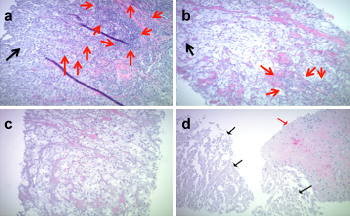
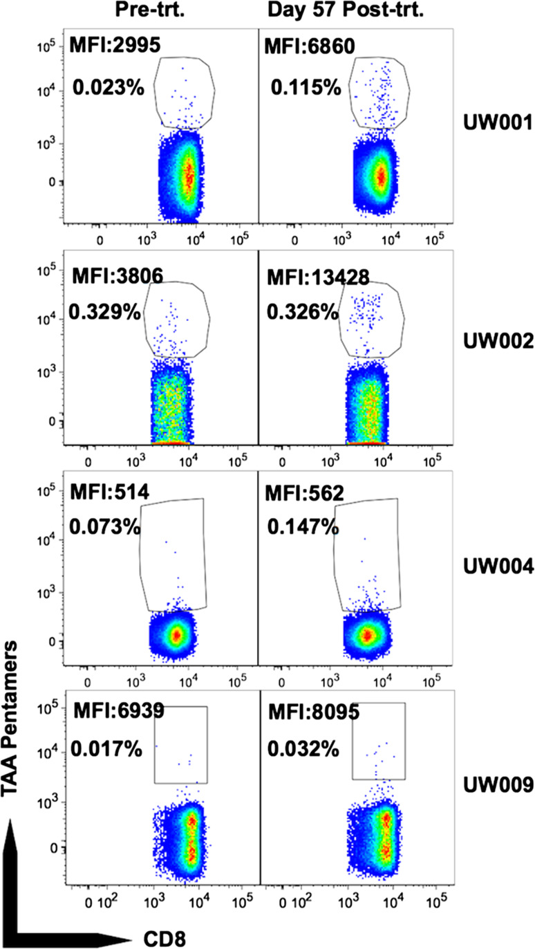
Effective uptake of tumor cell-derived antigens by antigen-presenting cells is achieved pre-clinically by in situ labeling of tumor with α-gal glycolipids that bind the naturally occurring anti-Gal antibody. We evaluated toxicity and feasibility of intratumoral injections of α-gal glycolipids as an autologous tumor antigen-targeted immunotherapy in melanoma patients (pts). Pts with unresectable metastatic melanoma, at least one cutaneous, subcutaneous, or palpable lymph node metastasis, and serum anti-Gal titer ≥1:50 were eligible for two intratumoral α-gal glycolipid injections given 4 weeks apart (cohort I: 0.1 mg/injection; cohort II: 1.0 mg/injection; cohort III: 10 mg/injection). Monitoring included blood for clinical, autoimmune, and immunological analyses and core tumor biopsies. Treatment outcome was determined 8 weeks after the first α-gal glycolipid injection. Nine pts received two intratumoral injections of α-gal glycolipids (3 pts/cohort). Injection-site toxicity was mild, and no systemic toxicity or autoimmunity could be attributed to the therapy. Two pts had stable disease by RECIST lasting 8 and 7 months. Tumor nodule biopsies revealed minimal to no change in inflammatory infiltrate between pre- and post-treatment biopsies except for 1 pt (cohort III) with a post-treatment inflammatory infiltrate. Two and four weeks post-injection, treated nodules in 5 of 9 pts exhibited tumor cell necrosis without neutrophilic or lymphocytic inflammatory response. Non-treated tumor nodules in 2 of 4 evaluable pts also showed necrosis. Repeated intratumoral injections of α-gal glycolipids are well tolerated, and tumor necrosis was seen in some tumor nodule biopsies after tumor injection with α-gal glycolipids.
Keywords: Cancer vaccines; Immunotherapy; Melanoma; α-Gal glycolipids.
Conflict of interest statement
The authors have the following financial or other conflicts of interests to disclose related to this publication: Uri Galili is the inventor of this immunotherapy and is a consultant to Agalimmune Inc. which further develops cancer immunotherapy with α-gal glycolipids. All other authors declare no financial or other conflicts of interests related to this publication.
Figures
Similar articles
-
Self-Tumor Antigens in Solid Tumors Turned into Vaccines by α-gal Micelle Immunotherapy.Pharmaceutics. 2024 Sep 27;16(10):1263. doi: 10.3390/pharmaceutics16101263. Pharmaceutics. 2024. PMID: 39458595 Free PMC article. Review.
-
Cancer immunotherapy by intratumoral injection of α-gal glycolipids.Anticancer Res. 2012 Sep;32(9):3861-8. Anticancer Res. 2012. PMID: 22993330 Clinical Trial.
-
Intratumoral injection of alpha-gal glycolipids induces xenograft-like destruction and conversion of lesions into endogenous vaccines.J Immunol. 2007 Apr 1;178(7):4676-87. doi: 10.4049/jimmunol.178.7.4676. J Immunol. 2007. PMID: 17372027
-
Conversion of tumors into autologous vaccines by intratumoral injection of α-Gal glycolipids that induce anti-Gal/α-Gal epitope interaction.Clin Dev Immunol. 2011;2011:134020. doi: 10.1155/2011/134020. Epub 2011 Nov 17. Clin Dev Immunol. 2011. PMID: 22162709 Free PMC article. Review.
-
Intratumoral injection of alpha-gal glycolipids induces a protective anti-tumor T cell response which overcomes Treg activity.Cancer Immunol Immunother. 2009 Oct;58(10):1545-56. doi: 10.1007/s00262-009-0662-2. Epub 2009 Jan 28. Cancer Immunol Immunother. 2009. PMID: 19184002 Free PMC article.
Cited by
-
Self-Tumor Antigens in Solid Tumors Turned into Vaccines by α-gal Micelle Immunotherapy.Pharmaceutics. 2024 Sep 27;16(10):1263. doi: 10.3390/pharmaceutics16101263. Pharmaceutics. 2024. PMID: 39458595 Free PMC article. Review.
-
AGI-134: a fully synthetic α-Gal glycolipid that converts tumors into in situ autologous vaccines, induces anti-tumor immunity and is synergistic with an anti-PD-1 antibody in mouse melanoma models.Cancer Cell Int. 2019 Dec 19;19:346. doi: 10.1186/s12935-019-1059-8. eCollection 2019. Cancer Cell Int. 2019. PMID: 31889898 Free PMC article.
-
Increasing Efficacy of Enveloped Whole-Virus Vaccines by In situ Immune-Complexing with the Natural Anti-Gal Antibody.Med Res Arch. 2021 Jul;9(7):2481. doi: 10.18103/mra.v9i7.2481. Epub 2021 Jul 10. Med Res Arch. 2021. PMID: 34853815 Free PMC article.
-
Tumor treatment by pHLIP-targeted antigen delivery.Front Bioeng Biotechnol. 2023 Jan 6;10:1082290. doi: 10.3389/fbioe.2022.1082290. eCollection 2022. Front Bioeng Biotechnol. 2023. PMID: 36686229 Free PMC article.
-
Heterosubtypic Protection Induced by a Live Attenuated Influenza Virus Vaccine Expressing Galactose-α-1,3-Galactose Epitopes in Infected Cells.mBio. 2020 Mar 3;11(2):e00027-20. doi: 10.1128/mBio.00027-20. mBio. 2020. PMID: 32127444 Free PMC article.
References
Publication types
MeSH terms
Substances
Grants and funding
LinkOut - more resources
Full Text Sources
Other Literature Sources
Medical
