

Role of Cdc6 in re-replication in cells expressing human papillomavirus E7 oncogene
- PMID: 27207654
- PMCID: PMC4967213
- DOI: 10.1093/carcin/bgw059
Role of Cdc6 in re-replication in cells expressing human papillomavirus E7 oncogene
Abstract
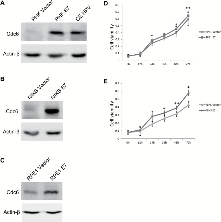
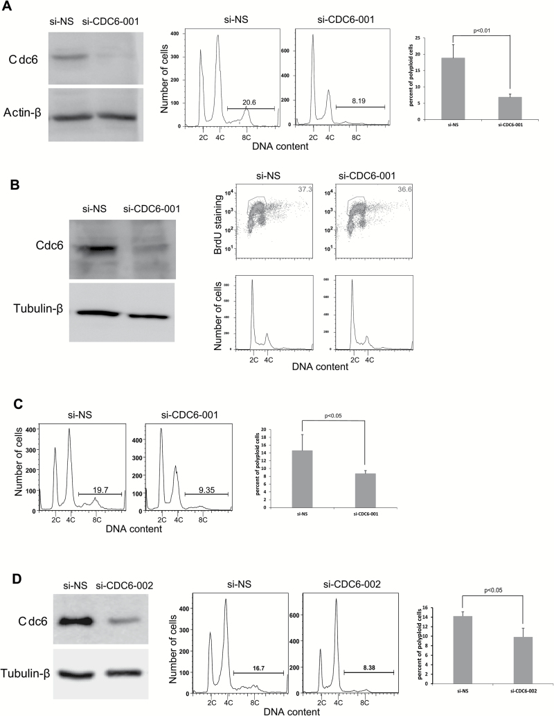
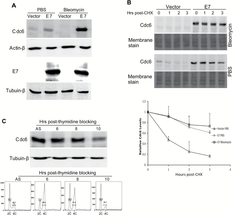
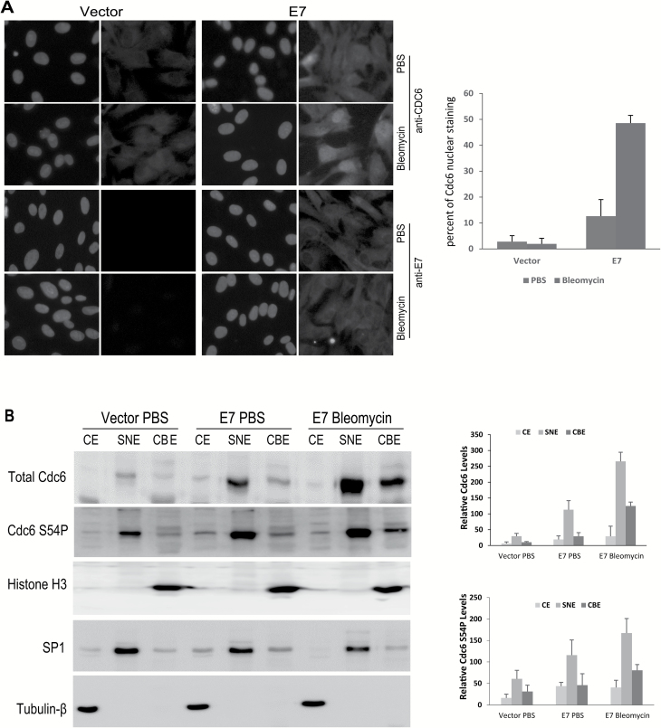
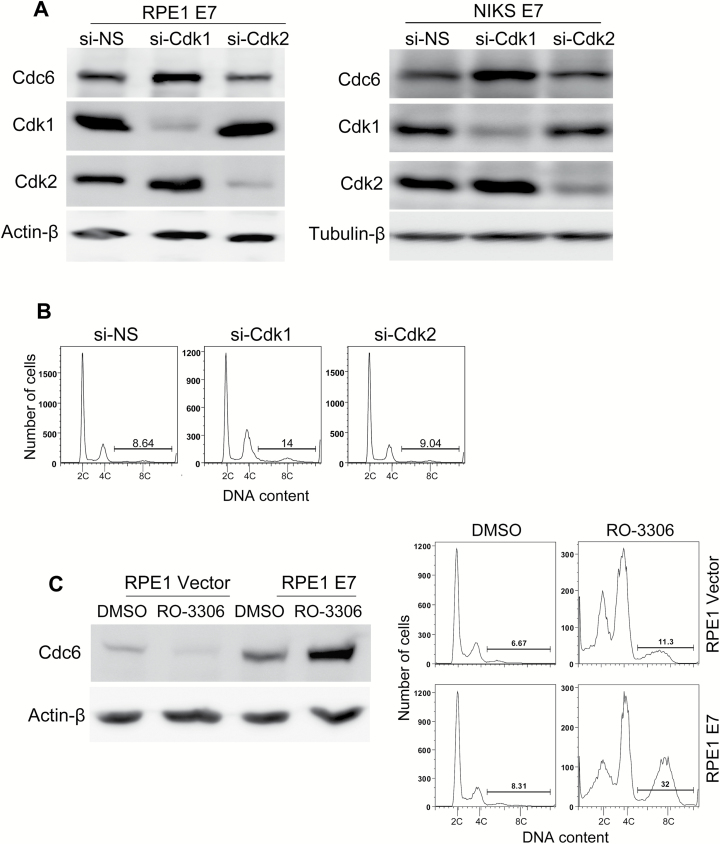
The E7 oncoprotein of high-risk human papillomavirus (HPV) types induces DNA re-replication that contributes to carcinogenesis; however, the mechanism is not fully understood. To better understand the mechanism by which E7 induces re-replication, we investigated the expression and function of cell division cycle 6 (Cdc6) in E7-expressing cells. Cdc6 is a DNA replication initiation factor and exhibits oncogenic activities when overexpressed. We found that in E7-expressing cells, the steady-state level of Cdc6 protein was upregulated and its half-life was increased. Cdc6 was localized to the nucleus and associated with chromatin, especially upon DNA damage. Importantly, downregulation of Cdc6 reduced E7-induced re-replication. Interestingly, the level of Cdc6 phosphorylation at serine 54 (S54P) was increased in E7-expressing cells. S54P was associated with an increase in the total amount of Cdc6 and chromatin-bound Cdc6. DNA damage-enhanced upregulation and chromatin binding of Cdc6 appeared to be due to downregulation of cyclin-dependent kinase 1 (Cdk1) as Cdk1 knockdown increased Cdc6 levels. Furthermore, Cdk1 knockdown or inhibition led to re-replication. These findings shed light on the mechanism by which HPV induces genomic instability and may help identify potential targets for drug development.
© The Author 2016. Published by Oxford University Press. All rights reserved. For Permissions, please email: journals.permissions@oup.com.
Figures
Similar articles
-
Role of WDHD1 in Human Papillomavirus-Mediated Oncogenesis Identified by Transcriptional Profiling of E7-Expressing Cells.J Virol. 2016 Jun 10;90(13):6071-6084. doi: 10.1128/JVI.00513-16. Print 2016 Jul 1. J Virol. 2016. PMID: 27099318 Free PMC article.
-
The lysine acetyltransferase GCN5 contributes to human papillomavirus oncoprotein E7-induced cell proliferation via up-regulating E2F1.J Cell Mol Med. 2018 Nov;22(11):5333-5345. doi: 10.1111/jcmm.13806. Epub 2018 Aug 6. J Cell Mol Med. 2018. PMID: 30079588 Free PMC article.
-
Role of Cdk1 in DNA damage-induced G1 checkpoint abrogation by the human papillomavirus E7 oncogene.Cell Cycle. 2014;13(20):3249-59. doi: 10.4161/15384101.2014.953879. Cell Cycle. 2014. PMID: 25485505 Free PMC article.
-
The human papillomavirus E7 oncoprotein as a regulator of transcription.Virus Res. 2017 Mar 2;231:56-75. doi: 10.1016/j.virusres.2016.10.017. Epub 2016 Nov 8. Virus Res. 2017. PMID: 27818212 Free PMC article. Review.
-
Cdc6 as a novel target in cancer: Oncogenic potential, senescence and subcellular localisation.Int J Cancer. 2020 Sep 15;147(6):1528-1534. doi: 10.1002/ijc.32900. Epub 2020 Feb 17. Int J Cancer. 2020. PMID: 32010971 Free PMC article. Review.
Cited by
-
Gene expression profile of THZ1-treated nasopharyngeal carcinoma cell lines indicates its involvement in the inhibition of the cell cycle.Transl Cancer Res. 2021 Jan;10(1):445-460. doi: 10.21037/tcr-19-2888. Transl Cancer Res. 2021. PMID: 35116274 Free PMC article.
-
The Not-So-Good, the Bad and the Ugly: HPV E5, E6 and E7 Oncoproteins in the Orchestration of Carcinogenesis.Viruses. 2021 Sep 22;13(10):1892. doi: 10.3390/v13101892. Viruses. 2021. PMID: 34696321 Free PMC article. Review.
-
Cdc6 contributes to abrogating the G1 checkpoint under hypoxic conditions in HPV E7 expressing cells.Sci Rep. 2017 Jun 7;7(1):2927. doi: 10.1038/s41598-017-03060-w. Sci Rep. 2017. PMID: 28592805 Free PMC article.
-
Polyploid Giant Cancer Cells, a Hallmark of Oncoviruses and a New Therapeutic Challenge.Front Oncol. 2020 Oct 14;10:567116. doi: 10.3389/fonc.2020.567116. eCollection 2020. Front Oncol. 2020. PMID: 33154944 Free PMC article. Review.
-
The prognostic significance of Cdc6 and Cdt1 in breast cancer.Sci Rep. 2017 Apr 20;7(1):985. doi: 10.1038/s41598-017-00998-9. Sci Rep. 2017. PMID: 28428557 Free PMC article.
References
-
- de Villiers E.M. (2013) Cross-roads in the classification of papillomaviruses. Virology, 445, 2–10. - PubMed
-
- Munoz N., et al. (2003) Epidemiologic classification of human papillomavirus types associated with cervical cancer. N. Engl. J. Med., 348, 518–527. - PubMed
-
- zur Hausen H. (2002) Papillomaviruses and cancer: from basic studies to clinical application. Nat. Rev. Cancer, 2, 342–350. - PubMed
-
- Walboomers J.M., et al. (1999) Human papillomavirus is a necessary cause of invasive cervical cancer worldwide. J. Pathol., 189, 12–19. - PubMed
Publication types
MeSH terms
Substances
Grants and funding
LinkOut - more resources
Full Text Sources
Other Literature Sources
Molecular Biology Databases
Miscellaneous
