

Patterns of expression of factor VIII and von Willebrand factor by endothelial cell subsets in vivo
- PMID: 27207787
- PMCID: PMC4937354
- DOI: 10.1182/blood-2015-12-684688
Patterns of expression of factor VIII and von Willebrand factor by endothelial cell subsets in vivo
Abstract
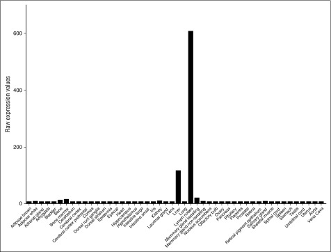
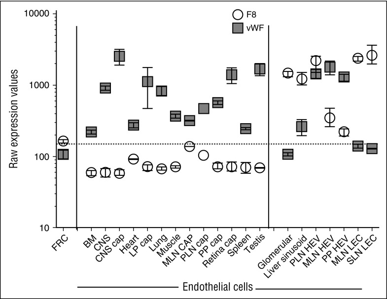
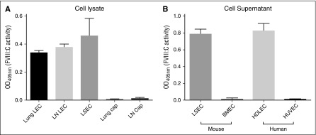
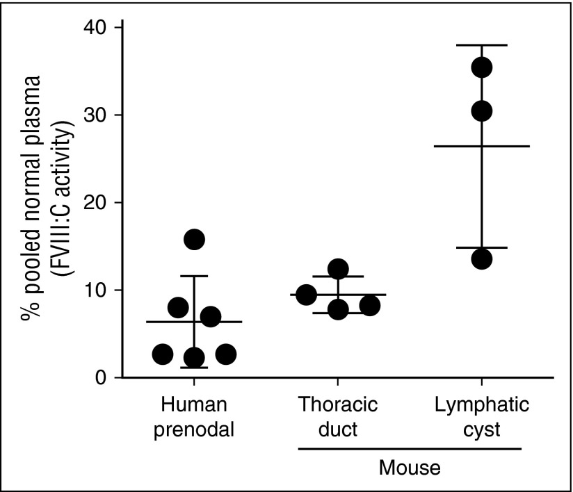
Circulating factor VIII (FVIII) is derived from liver and from extrahepatic sources probably of endothelial origin, but the vascular sites of FVIII production remain unclear. Among organs profiled, only liver and lymph nodes (LNs) show abundant expression of F8 messenger RNA (mRNA). Transcriptomic profiling of subsets of stromal cells, including endothelial cells (ECs) from mouse LNs and other tissues, showed that F8 mRNA is expressed by lymphatic ECs (LECs) but not by capillary ECs (capECs), fibroblastic reticular cells, or hematopoietic cells. Among blood ECs profiled, F8 expression was seen only in fenestrated ECs (liver sinusoidal and renal glomerular ECs) and some high endothelial venules. In contrast, von Willebrand factor mRNA was expressed in capECs but not in LECs; it was coexpressed with F8 mRNA in postcapillary high endothelial venules. Purified LECs and liver sinusoidal ECs but not capECs from LNs secrete active FVIII in culture, and human and mouse lymph contained substantial
Fviii: C activity. Our results revealed localized vascular expression of FVIII and von Willebrand factor and identified LECs as a major cellular source of FVIII in extrahepatic tissues.
Figures
Similar articles
-
Tissue distribution of factor VIII gene expression in vivo--a closer look.Thromb Haemost. 2001 Sep;86(3):855-61. Thromb Haemost. 2001. PMID: 11583319
-
Distribution of factor VIII mRNA and antigen in human liver and other tissues.Nature. 1985 Oct 24-30;317(6039):726-9. doi: 10.1038/317726a0. Nature. 1985. PMID: 3932885
-
Tissue distribution and regulation of murine von Willebrand factor gene expression in vivo.Blood. 1998 Oct 15;92(8):2791-801. Blood. 1998. PMID: 9763564
-
Pathophysiological Mechanisms of Endogenous FVIII Release following Strenuous Exercise in Non-severe Haemophilia: A Review.Thromb Haemost. 2017 Dec;117(12):2237-2242. doi: 10.1160/TH17-01-0004. Epub 2017 Dec 6. Thromb Haemost. 2017. PMID: 29212111 Review.
-
Von Willebrand Factor, Factor VIII, and Other Acute Phase Reactants as Biomarkers of Inflammation and Endothelial Dysfunction in Chronic Graft-Versus-Host Disease.Front Immunol. 2021 Apr 30;12:676756. doi: 10.3389/fimmu.2021.676756. eCollection 2021. Front Immunol. 2021. PMID: 33995421 Free PMC article. Review.
Cited by
-
Coagulation biomarkers are independent predictors of increased oxygen requirements in COVID-19.J Thromb Haemost. 2020 Nov;18(11):2942-2953. doi: 10.1111/jth.15067. Epub 2020 Sep 18. J Thromb Haemost. 2020. PMID: 32881304 Free PMC article.
-
Therapeutic potential of fetal liver cell transplantation in hemophilia A mice.Haematologica. 2023 Jun 1;108(6):1544-1554. doi: 10.3324/haematol.2022.282001. Haematologica. 2023. PMID: 36700401 Free PMC article.
-
Advances in Clinical and Basic Science of Coagulation: Illustrated abstracts of the 9th Chapel Hill Symposium on Hemostasis.Res Pract Thromb Haemost. 2018 Apr 12;2(3):407-428. doi: 10.1002/rth2.12095. eCollection 2018 Jul. Res Pract Thromb Haemost. 2018. PMID: 30046746 Free PMC article. Review.
-
The role of microRNAs in defining LSECs cellular identity and in regulating F8 gene expression.Front Genet. 2024 Feb 19;15:1302685. doi: 10.3389/fgene.2024.1302685. eCollection 2024. Front Genet. 2024. PMID: 38440189 Free PMC article.
-
Microvascular Thrombosis and Liver Fibrosis Progression: Mechanisms and Clinical Applications.Cells. 2023 Jun 24;12(13):1712. doi: 10.3390/cells12131712. Cells. 2023. PMID: 37443746 Free PMC article. Review.
References
-
- Vlot AJ, Koppelman SJ, van den Berg MH, Bouma BN, Sixma JJ. The affinity and stoichiometry of binding of human factor VIII to von Willebrand factor. Blood. 1995;85(11):3150–3157. - PubMed
-
- Tuddenham E. Far away and long ago. J Thromb Haemost. 2014;12(1):34–35. - PubMed
-
- Marchioro TL, Hougie C, Ragde H, Epstein RB, Thomas ED. Hemophilia: role of organ homografts. Science. 1969;163(3863):188–190. - PubMed
MeSH terms
Substances
Grants and funding
LinkOut - more resources
Full Text Sources
Other Literature Sources
Miscellaneous
