. 2016 Jul 14;128(2):249-52.
doi: 10.1182/blood-2016-03-704460. Epub 2016 May 18.

A clinically relevant in vivo zebrafish model of human multiple myeloma to study preclinical therapeutic efficacy

Affiliations

A clinically relevant in vivo zebrafish model of human multiple myeloma to study preclinical therapeutic efficacy

Jianhong Lin et al. Blood. .

Abstract

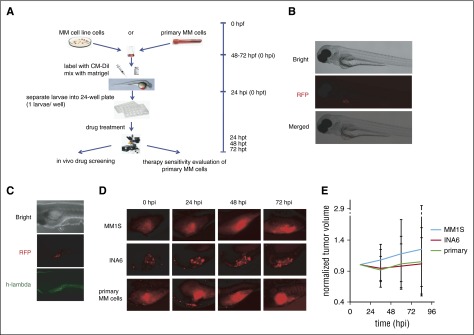
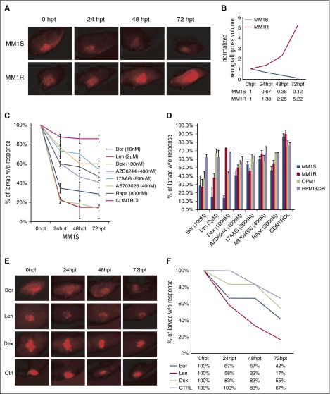
Patient-derived multiple myeloma (MM) cells are difficult to establish in culture or propagate in vivo in murine model. Here, we describe a zebrafish xenograft model that permits rapid, reliable growth of human MM cells injected into the perivitelline space of albino zebrafish (Casper) embryos 48 hours postfertilization. MM1S and MM1R MM cell lines and primary CD138(+) MM cells were stained with CM-Dil red fluorescent dye and suspended in Matrigel prior to their injection. The cells grew at the site of injection and disseminated throughout the developing embryos and larvae. Tumor size was quantified by fluorescent microscopy, and cell fate was followed for 4 days. All of the cell line xenografts showed responses similar to those previously observed with in vitro assays. CD138(+) plasma cell xenografts derived from MM patients also grew and were inhibited by the same drugs patients had responded to clinically. Using this technique, we can assess drug sensitivity or resistance with a small number of MM cells in a short period. This raises the possibility that one might be able to assess drug sensitivity in real time with readily obtainable clinical samples.


Figures

Figure 1
Figure 1
Casper zebrafish support MM cell growth. (A) Schematic chart depicting the procedures used to establish the zebrafish MM xenograft model (hpf, hours postfertilization; hpi, hours postinjection; hpt, hours posttreatment). (B) Fluorescence microscopy images of the CM-Dil–stained MM cells injected into the perivitelline area of Casper zebrafish larvae. Bright field of whole Casper zebrafish 24 hours after injection with human MM cells (top). Red fluorescence protein (RFP) channel of whole Casper zebrafish; the red fluorescence color under the microscope shows CM-Dil–stained MM cells (middle). Merged image from bright field and RFP field (bottom). All cells are located in the perivitelline area after mixing with Matrigel. (C) Whole-mount immunohistochemistry staining. Bright field of whole zebrafish 24 hpi with human multiple MM cells (top). RFP field shows red fluorescence from CM-Dil–stained MM cells (middle). GFP field showing the EGFP-labeled anti–human λ light-chain antibody colocalizing with CM-Dil–labeled MM1S cells (bottom). (D) Typical fluorescence microscopy images of the CM-Dil–stained MM cells xenograft growth of MM1S, INA-6, and primary patient MM cells without drug treatment in the perivitelline area of Casper zebrafish larvae at 24, 48, and 72 hpi. (E) Tumor growth curves of xenografts with these 3 cell types in Casper zebrafish larvae (n = 30 for each group).
Figure 2
Figure 2
Response to standard and novel drugs in the zebrafish model. (A) Fluorescent microscopy images of CM-Dil–stained MM1S and MM1R xenografts at 0, 24, 48, and 72 hpi. (B) Tumor growth curves of MM1S and MM1R cells xenografts indicating that MM1S xenografts are responsive to dexamethasone treatment whereas MM1R xenografts are resistant. (C) Percentage of MM1S xenografts in zebrafish showing no response after treatment with bortezomib (Bor; n = 22), lenalidomide (Len; n = 22), dexamethasone (Dex; n = 22), AZD6244 (n = 18), 17-AAG (n = 22), rapamycin (Rapa; n = 18), AS703026 (n = 18), and dimethyl sulfoxide (control; n = 20), respectively. Error bars indicate standard errors. (D) Summary of percentage of MM1S, MM1R, OPM1, and RPMI8226 larvae without response with various indicated novel agents. (E) Individual fluorescence microscopy images of CD138+ MM cells from newly diagnosed MM patient xenografts in zebrafish before and after treatment with bortezomib (n = 8), lenalidomide (n = 8), dexamethasone (n = 8), and dimethyl sulfoxide (n = 8), at 0, 24, 48, and 72 hpt, respectively. (F) Percentage of primary patient MM cell xenografts without response to lenalidomide, bortezomib, and dexamethasone. Ctrl, control.

References

    1. Amin SB, Yip WK, Minvielle S, et al. Gene expression profile alone is inadequate in predicting complete response in multiple myeloma. Leukemia. 2014;28(11):2229–2234. - PMC - PubMed
    1. Carrasco DR, Sukhdeo K, Protopopova M, et al. The differentiation and stress response factor XBP-1 drives multiple myeloma pathogenesis. Cancer Cell. 2007;11(4):349–360. - PMC - PubMed
    1. Chesi M, Robbiani DF, Sebag M, et al. AID-dependent activation of a MYC transgene induces multiple myeloma in a conditional mouse model of post-germinal center malignancies. Cancer Cell. 2008;13(2):167–180. - PMC - PubMed
    1. Groen RW, Noort WA, Raymakers RA, et al. Reconstructing the human hematopoietic niche in immunodeficient mice: opportunities for studying primary multiple myeloma. Blood. 2012;120(3):e9–e16. - PubMed
    1. Rongvaux A, Willinger T, Martinek J, et al. Development and function of human innate immune cells in a humanized mouse model. Nat Biotechnol. 2014;32(4):364–372. - PMC - PubMed

Publication types