

Induction of matrix metalloproteinase-1 by tumor necrosis factor-α is mediated by interleukin-6 in cultured fibroblasts of keratoconus
- PMID: 27207902
- PMCID: PMC5102130
- DOI: 10.1177/1535370216650940
Induction of matrix metalloproteinase-1 by tumor necrosis factor-α is mediated by interleukin-6 in cultured fibroblasts of keratoconus
Abstract
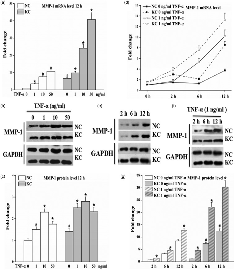
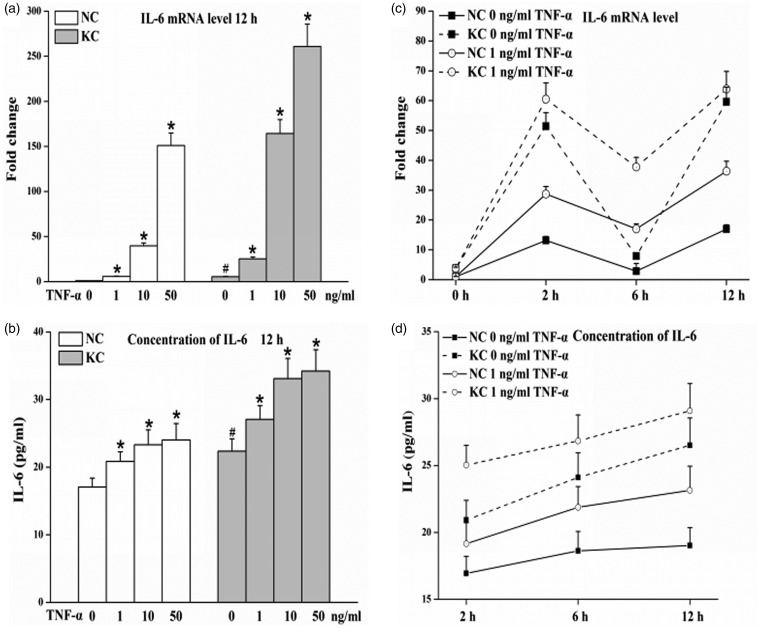
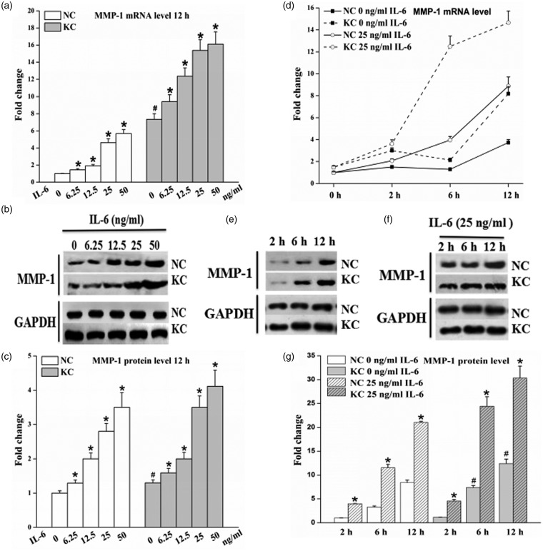
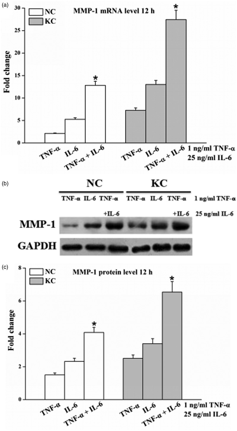
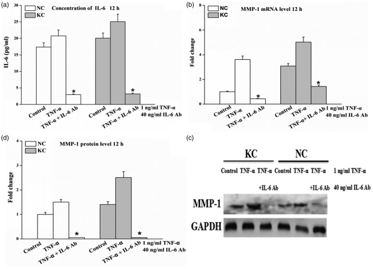
Inflammatory molecules and matrix metalloproteinase (MMPs) have been found over-expressed in the tear film of patients with keratoconus. However, the mechanistic link between inflammatory molecules and MMPs in the pathogenesis of keratoconus remains still elusive. Therefore, we investigated the effect of tumor necrosis factor-α (TNF-α) and interleukin-6 (IL-6) on MMP-1 expression and used IL-6 antibody (IL-6 Ab) to examine the role of IL-6 on TNF-α mediated regulation of MMP-1 in fibroblasts of normal cornea and keratoconus. Real-time polymerase chain reaction, Enzyme-linked immunosorbent assay, and Western blot data demonstrated that MMP-1 and IL-6 were expressed in fibroblasts of normal cornea and keratoconus. Levels of MMP-1 and IL-6 were significantly higher in keratoconus than normal cornea. TNF-α treatment led to a significant increase in IL-6 levels. IL-6 treatment induced MMP-1 synthesis in normal cornea and keratoconus. TNF-α increased MMP-1 expression in a dose- and time-dependent manner and this response was completely inhibited by the IL-6 Ab. In conclusion, these results indicate that fibroblasts of keratoconus shows increased levels of IL-6 and MMP-1 gene and protein expression and IL-6 mediates the TNF-α-induced MMP-1 expression.
Keywords: Corneal fibroblasts; inflammatory cytokine; keratoconus; matrix metalloproteinases (MMPs).
Figures
Similar articles
-
[Effects of Tumor Necrosis Factor Alpha on the Expression of Matrix Metalloproteinases and Tissue Inhibitors of Matrix Metalloproteinases in Keratoconus Fibroblasts].Sheng Wu Yi Xue Gong Cheng Xue Za Zhi. 2016 Dec;33(6):1139-44. Sheng Wu Yi Xue Gong Cheng Xue Za Zhi. 2016. PMID: 29714979 Chinese.
-
Chronic exposure of interleukin-13 suppress the induction of matrix metalloproteinase-1 by tumour necrosis factor α in normal and scleroderma dermal fibroblasts through protein kinase B/Akt.Clin Exp Immunol. 2018 Jan;191(1):84-95. doi: 10.1111/cei.13045. Epub 2017 Oct 2. Clin Exp Immunol. 2018. PMID: 28884475 Free PMC article.
-
Influence of cytokines on matrix metalloproteinases produced by fibroblasts cultured in monolayer and collagen gels.J Formos Med Assoc. 2001 Jun;100(6):377-82. J Formos Med Assoc. 2001. PMID: 11480246
-
Keratoconus: an inflammatory disorder?Eye (Lond). 2015 Jul;29(7):843-59. doi: 10.1038/eye.2015.63. Epub 2015 May 1. Eye (Lond). 2015. PMID: 25931166 Free PMC article. Review.
-
Matrix metalloproteinases in keratoconus - Too much of a good thing?Exp Eye Res. 2019 May;182:137-143. doi: 10.1016/j.exer.2019.03.016. Epub 2019 Mar 23. Exp Eye Res. 2019. PMID: 30910610 Review.
Cited by
-
Exopolysaccharide from Lactobacillus plantarum HY7714 Protects against Skin Aging through Skin-Gut Axis Communication.Molecules. 2021 Mar 16;26(6):1651. doi: 10.3390/molecules26061651. Molecules. 2021. PMID: 33809637 Free PMC article.
-
Systematically Displaying the Pathogenesis of Keratoconus via Multi-Level Related Gene Enrichment-Based Review.Front Med (Lausanne). 2022 Jan 24;8:770138. doi: 10.3389/fmed.2021.770138. eCollection 2021. Front Med (Lausanne). 2022. PMID: 35141241 Free PMC article.
-
De novo mutations of TUBA3D are associated with keratoconus.Sci Rep. 2017 Oct 19;7(1):13570. doi: 10.1038/s41598-017-13162-0. Sci Rep. 2017. PMID: 29051577 Free PMC article.
-
Histopathologic findings of keratoconus corneas underwent penetrating keratoplasty according to topographic measurements and keratoconus severity.Int J Ophthalmol. 2017 Nov 18;10(11):1640-1646. doi: 10.18240/ijo.2017.11.02. eCollection 2017. Int J Ophthalmol. 2017. PMID: 29181305 Free PMC article.
-
Skullcapflavone II Inhibits Degradation of Type I Collagen by Suppressing MMP-1 Transcription in Human Skin Fibroblasts.Int J Mol Sci. 2019 Jun 4;20(11):2734. doi: 10.3390/ijms20112734. Int J Mol Sci. 2019. PMID: 31167359 Free PMC article.
References
-
- Rabinowitz YS. Keratoconus. Surv Ophthalmol 1998; 42: 297–319. - PubMed
-
- Lema I, Durán JA. Inflammatory molecules in the tears of patients with keratoconus. Ophthalmology 2005; 112: 654–9. - PubMed
-
- Lema I, Sobrino T, Duran JA, Brea D, Diez-Feijoo E. Subclinical keratoconus and inflammatory molecules from tears. Br J Ophthalmol 2009; 93: 820–4. - PubMed
-
- McMonnies CW. Inflammation and keratoconus. Optom Vis Sci 2015; 92: e35–41. - PubMed
Publication types
MeSH terms
Substances
LinkOut - more resources
Full Text Sources
Other Literature Sources