. 2016 Oct;65(10):1681-9.
doi: 10.1136/gutjnl-2015-311004. Epub 2016 May 20.

Primary sclerosing cholangitis is characterised by intestinal dysbiosis independent from IBD

Affiliations

Primary sclerosing cholangitis is characterised by intestinal dysbiosis independent from IBD

João Sabino et al. Gut. 2016 Oct.

Abstract

Objective: Primary sclerosing cholangitis (PSC) is a chronic cholestatic liver disease often leading to end-stage liver disease. Its pathogenesis remains largely unknown, although frequent concomitant IBD hints towards common factors underlying gut and bile duct inflammation. Considering the mounting evidence on the involvement of the intestinal microbiota in initiating and determining IBD phenotype, we investigated intestinal microbiota composition in patients with PSC.

Design: Stool samples were collected from 147 individuals (52 patients with PSC, 52 age, gender and body mass index-matched healthy volunteers, 13 UC and 30 patients with Crohn's disease). An independent validation cohort of 14 PSC and 14 matched controls was recruited. 16S rDNA sequencing of faecal DNA was performed (Illumina MiSeq).

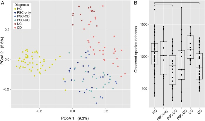
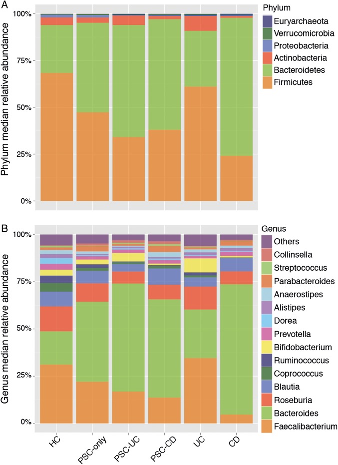
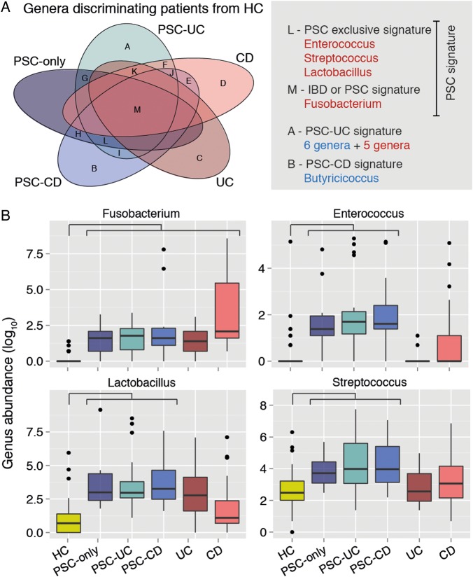
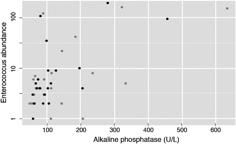
Results: The microbiota of patients with PSC was characterised by decreased microbiota diversity, and a significant overrepresentation of Enterococcus (p=3.76e-05), Fusobacterium (p=3.76e-05) and Lactobacillus (p=0.0002) genera. This dysbiosis was present in patients with PSC with and without concomitant IBD and was distinct from IBD, and independent of treatment with ursodeoxycholic acid. A decision tree based on abundances of these three genera allowed reliable classification in the validation cohort. In particular, one operational taxonomic unit belonging to the Enterococcus genus was associated with increased levels of serum alkaline phosphatase (p=0.048), a marker of disease severity.

Conclusions: We here present the first report of PSC-associated faecal dysbiosis, independent from IBD signatures, suggesting the intestinal microbiota could be a contributing factor in PSC pathogenesis. Further studies are needed to confirm these findings and assess causality.

Keywords: INTESTINAL MICROBIOLOGY; PRIMARY SCLEROSING CHOLANGITIS.


Figures

Figure 1
Figure 1
Faecal microbiota variation across healthy controls (HC) and patients with primary sclerosing cholangitis (PSC) and IBD (n=147). (A) Variation in microbial community composition represented in a principal coordinates analysis (PCoA) of the Bray–Curtis dissimilarity matrix, calculated from the operational taxonomic unit (OTU)-level abundance matrix. Patients with HC, PSC and IBD are significantly different (see online supplementary table S2), while PSC subgroups are not. (B) Box plots representation of microbiota richness (number of observed OTUs per sample) distribution across HC, PSC (PSC only, PSC-ulcerative colitis (UC) and PSC-Crohn's disease (CD)) and IBD (UC and CD). HC have significantly higher species richness than all patients with PSC combined, and than PSC-UC and CD individually. Horizontal bars represent false discovery rate (FDR)-corrected p value <0.05 (Kruskal–Wallis rank sum test). The body of the box plot represents the first and third quartiles of the distribution, and the median. The whiskers extend from the quartiles to the last datapoint within 1.5× IQR, with outliers beyond represented as dots.
Figure 2
Figure 2
Faecal microbiota composition in healthy controls, patients with primary sclerosing cholangitis (PSC) (PSC-only, PSC-ulcerative colitis (UC) and PSC-Crohn's disease (CD)) and IBD (UC and CD) (n=147). (A) Phylum-level median relative abundances and (B) genus-level median relative abundances. The top 14 more abundant genera are represented and all others are summed into the category ‘others’.
Figure 3
Figure 3
Genus-level microbiota signature of primary sclerosing cholangitis (PSC) (n=147). (A) Venn diagram summarising the overlap between the lists of genera with significantly different abundances in patient groups compared with healthy controls (HC). Genera overrepresented in patients as compared with controls are in red, and underrepresented ones in blue. The lists of genera included in each of the Venn diagram's sets (labelled from A to M) can be found in online supplementary table S3. (B) Box plots showing abundances of Fusobacterium, Enterococcus, Lactobacillus and Streptococcus in HC, patients with PSC and IBD. These compose the PSC signature (ie, genera significantly overrepresented or underrepresented in all PSC subgroups compared with HC), with the exception of Streptococcus, which association falls below significance after excluding patients under antibiotic treatment. Horizontal bar represents false discovery rate (FDR)-corrected p value <0.05 (Mann–Whitney test). Box plot definition is provided in the legend of figure 1.
Figure 4
Figure 4
Positive correlation between Enterococcus abundance and alkaline phosphatase (ALP), a clinical marker for cholestasis and primary sclerosing cholangitis (PSC) progression (n=87, Spearman ρ=0.29, adjusted p value=0.012199). Black dots correspond to patients with PSC without cirrhosis or liver transplantation. Grey dots correspond to patients with PSC with either cirrhosis or liver transplantation.

Comment in

Similar articles

Cited by

References

    1. Hirschfield GM, Karlsen TH, Lindor KD, et al. . Primary sclerosing cholangitis. Lancet 2013;382:1587–99. 10.1016/S0140-6736(13)60096-3 - DOI - PubMed
    1. Alabraba E, Nightingale P, Gunson B, et al. . A re-evaluation of the risk factors for the recurrence of primary sclerosing cholangitis in liver allografts. Liver Transpl 2009;15:330–40. 10.1002/lt.21679 - DOI - PubMed
    1. Olsson R, Björnsson E, Bäckman L, et al. . Bile duct bacterial isolates in primary sclerosing cholangitis: a study of explanted livers. J Hepatol 1998;28:426–32. 10.1016/S0168-8278(98)80316-4 - DOI - PubMed
    1. Färkkilä M, Karvonen AL, Nurmi H, et al. . Metronidazole and ursodeoxycholic acid for primary sclerosing cholangitis: a randomized placebo-controlled trial. Hepatology 2004;40:1379–86. 10.1002/hep.20457 - DOI - PubMed
    1. Seidel D, Eickmeier I, Kühl AA, et al. . CD8T cells primed in the gut-associated lymphoid tissue induce immune-mediated cholangitis in mice. Hepatology 2014;59:601–11. 10.1002/hep.26702 - DOI - PubMed

Publication types

MeSH terms