

The Mouse Fetal Ovary Has Greater Sensitivity Than the Fetal Testis to Benzo[a]pyrene-Induced Germ Cell Death
- PMID: 27208085
- PMCID: PMC4960906
- DOI: 10.1093/toxsci/kfw083
The Mouse Fetal Ovary Has Greater Sensitivity Than the Fetal Testis to Benzo[a]pyrene-Induced Germ Cell Death
Abstract
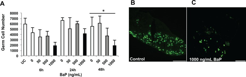
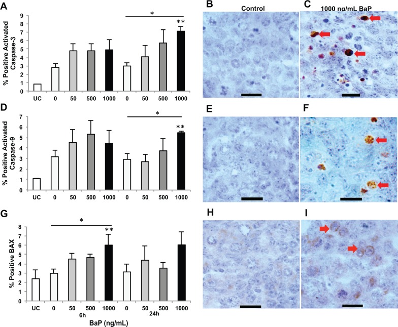
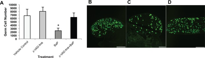
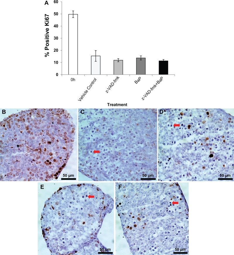
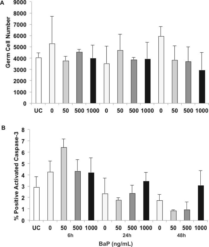
The polycyclic aromatic hydrocarbon pollutant benzo[a]pyrene (BaP) is a known developmental gonadotoxicant. However, the mechanism of BaP-induced germ cell death is unclear. We investigated whether exposure to BaP induces apoptotic germ cell death in the mouse fetal ovary or testis. Mouse fetal gonads were dissected at embryonic day 13.5 days postcoitum (dpc) and fixed immediately or cultured for 6, 24, 48, or 72 h with various concentrations of BaP (1-1000 ng/ml). Germ cells numbers, apoptosis, and proliferation were evaluated by immunostaining. Treatment of fetal ovaries with BaP for 72 h concentration-dependently depleted germ cells. Treatment with BaP elevated the expression of BAX protein at 6 h and activated downstream caspases-9 and -3 at 24 h in a concentration-dependent manner in germ cells of fetal ovaries. As a consequence, ovarian germ cell numbers were significantly and concentration-dependently decreased at 48 h. Pretreatment with z-VAD-fmk, a pan-caspase inhibitor, prior to exposure to 1000 ng/ml BaP prevented BaP-mediated ovarian germ cell death; there were no effects of BaP or z-VAD-fmk on germ cell proliferation. No significant effects of BaP exposure on caspase 3 activation or germ cell numbers were observed in fetal testes after 48 h of culture. Our findings show that BaP exposure increases caspase-dependent and BAX-associated germ cell apoptosis in the mouse fetal ovary, leading to germ cell depletion. In contrast, the cultured 13.5 dpc fetal testis is relatively resistant to BaP-induced germ cell death. This study provides a novel insight into molecular mechanisms by which BaP has direct gonadotoxicity in the mouse fetal ovary.
Keywords: apoptosis; benzo[a]pyrene; germ cells; ovary; polycyclic aromatic hydrocarbon; testis.
© The Author 2016. Published by Oxford University Press on behalf of the Society of Toxicology. All rights reserved. For Permissions, please e-mail: journals.permissions@oup.com.
Figures
Similar articles
-
Glutathione deficiency sensitizes cultured embryonic mouse ovaries to benzo[a]pyrene-induced germ cell apoptosis.Toxicol Appl Pharmacol. 2018 Aug 1;352:38-45. doi: 10.1016/j.taap.2018.05.024. Epub 2018 May 22. Toxicol Appl Pharmacol. 2018. PMID: 29800640 Free PMC article.
-
Sex Differences in Embryonic Gonad Transcriptomes and Benzo[a]pyrene Metabolite Levels After Transplacental Exposure.Endocrinology. 2022 Jan 1;163(1):bqab228. doi: 10.1210/endocr/bqab228. Endocrinology. 2022. PMID: 34734245 Free PMC article.
-
Gestational Benzo[a]pyrene Exposure Destroys F1 Ovarian Germ Cells Through Mitochondrial Apoptosis Pathway and Diminishes Surviving Oocyte Quality.Toxicol Sci. 2022 Oct 27;190(1):23-40. doi: 10.1093/toxsci/kfac086. Toxicol Sci. 2022. PMID: 35993611 Free PMC article.
-
Caspase-mediated apoptosis in the vertebrate ovary.Reproduction. 2002 Jul;124(1):19-27. doi: 10.1530/rep.0.1240019. Reproduction. 2002. PMID: 12090914 Review.
-
Embryology of the gonad with reference to special tumors of the ovary and testis.J Pediatr Surg. 1988 Oct;23(10):967-72. doi: 10.1016/s0022-3468(88)80396-8. J Pediatr Surg. 1988. PMID: 3069998 Review.
Cited by
-
Glutathione deficiency sensitizes cultured embryonic mouse ovaries to benzo[a]pyrene-induced germ cell apoptosis.Toxicol Appl Pharmacol. 2018 Aug 1;352:38-45. doi: 10.1016/j.taap.2018.05.024. Epub 2018 May 22. Toxicol Appl Pharmacol. 2018. PMID: 29800640 Free PMC article.
-
In Utero Exposure to Benzo[a]pyrene Induces Ovarian Mutations at Doses That Deplete Ovarian Follicles in Mice.Environ Mol Mutagen. 2019 Jun;60(5):410-420. doi: 10.1002/em.22261. Epub 2018 Dec 21. Environ Mol Mutagen. 2019. PMID: 30353947 Free PMC article.
-
Sex Differences in Embryonic Gonad Transcriptomes and Benzo[a]pyrene Metabolite Levels After Transplacental Exposure.Endocrinology. 2022 Jan 1;163(1):bqab228. doi: 10.1210/endocr/bqab228. Endocrinology. 2022. PMID: 34734245 Free PMC article.
-
Polycyclic Aromatic Hydrocarbons (PAHs) in the Environment: Occupational Exposure, Health Risks and Fertility Implications.Toxics. 2025 Feb 23;13(3):151. doi: 10.3390/toxics13030151. Toxics. 2025. PMID: 40137477 Free PMC article. Review.
-
Prenatal exposure to benzo[a]pyrene depletes ovarian reserve and masculinizes embryonic ovarian germ cell transcriptome transgenerationally.Sci Rep. 2023 May 29;13(1):8671. doi: 10.1038/s41598-023-35494-w. Sci Rep. 2023. PMID: 37248279 Free PMC article.
References
-
- Alton M., Taketo T. (2007). Switch from BAX-dependent to BAX-independent germ cell loss during the development of fetal mouse ovaries. J. Cell Sci. 130, 417–424. - PubMed
-
- Atchison F. W., Capel B., Means A. R. (2003). Pin1 regulates the timing of mammalian primordial germ cell proliferation. Development 130, 3579–3586. - PubMed
-
- ATSDR (1995). Toxicological Profile for Polycyclic Aromatic Hydrocarbons. US Department of Health and Human Services, Public Health Service, Agency for Toxic Substances and Disease Registry, Atlanta, GA. - PubMed
-
- Baltus A. E., Menke D. B., Hu Y. C., Goodheart M. L., Carpenter A. E., De Rooij D. G., Page D. C. (2006). In germ cells of mouse embryonic ovaries, the decision to enter meiosis precedes premeiotic DNA replication. Nat. Genet. 38, 1430–1434. - PubMed
Publication types
MeSH terms
Substances
Grants and funding
LinkOut - more resources
Full Text Sources
Other Literature Sources
Molecular Biology Databases
Research Materials
