. 2016 Jun;171(2):821-32.
doi: 10.1104/pp.15.01878. Epub 2016 Apr 4.

A Light Switch Based on Protein S-Nitrosylation Fine-Tunes Photosynthetic Light Harvesting in Chlamydomonas

Affiliations

A Light Switch Based on Protein S-Nitrosylation Fine-Tunes Photosynthetic Light Harvesting in Chlamydomonas

Hanna Berger et al. Plant Physiol. 2016 Jun.

Abstract

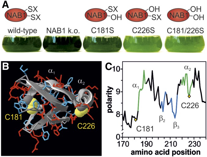
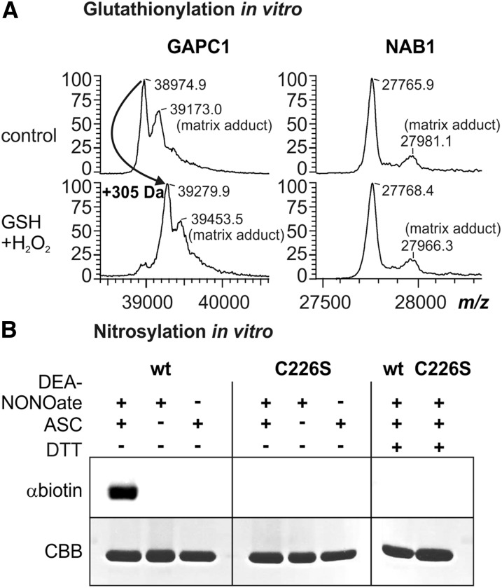
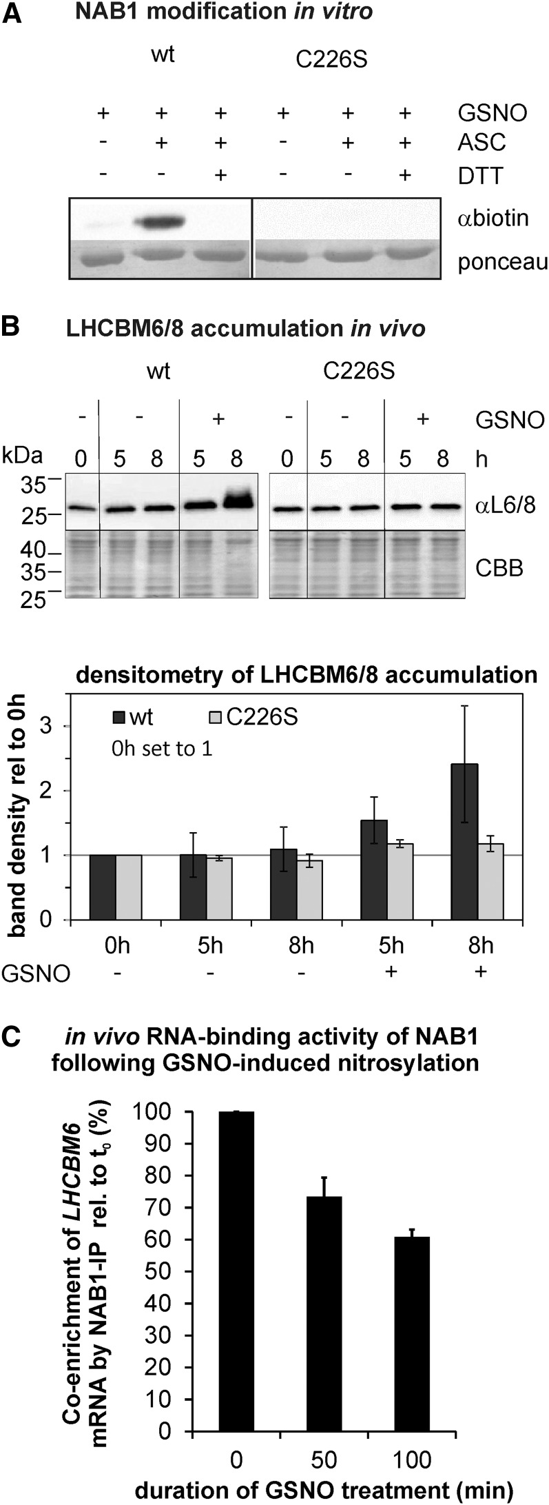
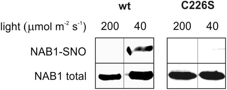
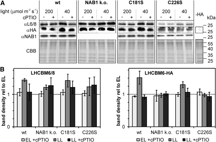
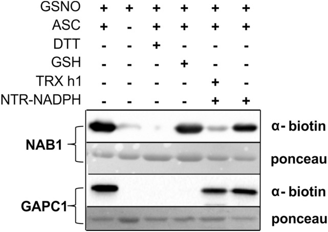
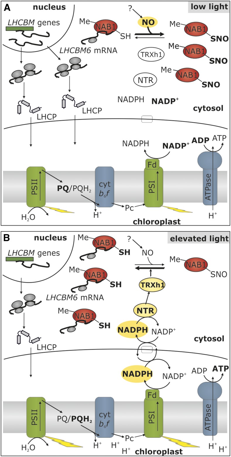
Photosynthetic eukaryotes are challenged by a fluctuating light supply, demanding for a modulated expression of nucleus-encoded light-harvesting proteins associated with photosystem II (LHCII) to adjust light-harvesting capacity to the prevailing light conditions. Here, we provide clear evidence for a regulatory circuit that controls cytosolic LHCII translation in response to light quantity changes. In the green unicellular alga Chlamydomonas reinhardtii, the cytosolic RNA-binding protein NAB1 represses translation of certain LHCII isoform mRNAs. Specific nitrosylation of Cys-226 decreases NAB1 activity and could be demonstrated in vitro and in vivo. The less active, nitrosylated form of NAB1 is found in cells acclimated to limiting light supply, which permits accumulation of light-harvesting proteins and efficient light capture. In contrast, elevated light supply causes its denitrosylation, thereby activating the repression of light-harvesting protein synthesis, which is needed to control excitation pressure at photosystem II. Denitrosylation of recombinant NAB1 is efficiently performed by the cytosolic thioredoxin system in vitro. To our knowledge, NAB1 is the first example of stimulus-induced denitrosylation in the context of photosynthetic acclimation. By identifying this novel redox cross-talk pathway between chloroplast and cytosol, we add a new key element required for drawing a precise blue print of the regulatory network of light harvesting.


Figures

Figure 1.
Figure 1.
Essentiality of Cys-226 for NAB1 redox control and in silico indication for its nitrosylation. A, Pale-green phenotype displayed by cell lines expressing NAB1 variants that lack Cys-226, which is essential for Cys-based redox control of its repressor activity. In these variants, cysteines amenable to redox-based modifications (-SX) are replaced with nonreactive serines (-OH) that mimic the free thiol state (-SH). B, In silico model of the NAB1-RRM domain. Polar and charged amino acids in the surrounding of cysteines 181 and 226 are shown in red, and uncharged/nonpolar amino acids are depicted in blue. The protein backbone is shown in a ribbon presentation. C, Polarity plot of the RRM domain. The relative polarity is shown on the y axis, while the amino acid position is given on the x axis. α-helices, β-sheets, and loop regions are indicated by a green, blue, and black color, respectively.
Figure 2.
Figure 2.
NAB1 can be nitrosylated in vitro. A, Analysis of NAB1 and GAPC1 in vitro glutathionylation following treatment with hydrogen peroxide (0.1 mm) and glutathione (0.5 mm) via MALDI-TOF mass spectrometry. Left panel: Mass spectrum of GAPC1 obtained after treatment with GSH+H2O2 and subsequent reduction using DTT (2.5 mm; used as a control). A mass increase of 305 D corresponds to one glutathione molecule covalently bound per protein monomer. The peaks labeled “matrix adducts” correspond to proteins with a sinapinic acid adduct. Differences between mass peaks of unmodified NAB1 and GAPC1 are within the experimental error of the instrument. Right panel: Mass spectrometric analysis of recombinant NAB1 under identical conditions. B, Treatment of recombinant NAB1 (wt) and NAB1C226S (C226S) with the NO-donor DEA-NONOate followed by the biotin switch technique. Addition (+) or omission (−) of the reaction components DEA-NONOate (1 mm), ascorbate (ASC; 40 mm), and DTT (20 mm) during the assay is indicated in the upper part. NAB1-biotinylation as an indicator for prior nitrosylation was detected by immunoblotting with a biotin-specific antiserum (αbiotin), and NAB1 protein amounts were assessed by Coomassie staining (CBB) after SDS-PAGE separation.
Figure 3.
Figure 3.
Nitrosylation of NAB1 at Cys-226 decreases its translation repressor activity. A, In vitro nitrosylation of recombinant NAB1 with GSNO (2 mm). The wild-type version of NAB1 was used along with NAB1C226S. The biotin switch technique was applied to detect S-nitrosylation via immunodetection of the biotin label (αbiotin). Omission of GSNO (−) and addition of DTT (+DTT) served as a control to assess stringency of the assay. Ponceau staining served as loading control. B, Effects of Cys-226 nitrosylation on the accumulation of major light-harvesting proteins LHCBM6/8. The cellular amount of LHCBM6/8 was determined by immunodetection (αL6/8; upper panel) 5 and 8 h following GSNO addition (+) to cultures expressing either wt-NAB1 (wt) or NAB1C226S (C226S) under control of the PSAD promoter. Negative controls (−GSNO) were included to exclude effects unrelated to nitrosative stress. Changes in the level of LHCBM6/8 relative to the t0 level of the wild type (black bars) or C226S (gray bars) were quantified by densitometric scanning (lower panel). Mean values from two biological replicates including two technical replicates are given along with standard deviations (n = 4). C, Effects of Cys-226 nitrosylation on the RNA-binding activity of NAB1 in vivo. Cys nitrosylation was induced by addition of GSNO to liquid cultures expressing wt-NAB1 under control of the PSAD promoter, and samples for the coimmunoprecipitation of LHCBM6-mRNA via NAB1 were taken at indicated time points (x axis). Enrichment factors for LHCBM6-mRNA were determined by comparing the relative LHCBM6 amount in input and coprecipitated samples for each time point by applying quantitative RT-PCR. The GSNO-induced change in LHCBM6-mRNA enrichment during NAB1 immunoprecipitation was calculated by setting the enrichment factor obtained before (t0min) GSNO addition to 100%. Error bars represent standard errors derived from four technical replicates (n = 4).
Figure 4.
Figure 4.
NAB1 is nitrosylated in vivo at Cys-226 under low-light conditions. C. reinhardtii cells overexpressing wt-NAB1 or NAB1C226S were grown under low light (40 μmol m−2 s−1) or elevated light (200 μmol m−2 s−1) for 8 h and then subjected to BST. After BST, biotinylated proteins were purified by streptavidin affinity chromatography and eluted with DTT. Eluted proteins were analyzed by western blot using αNAB1 antibodies (NAB1-SNO). Immunoblot detection of NAB1 before chromatography (NAB1 total) served as control. Bands shown in one panel derive from the same blot and were rearranged for clarity.
Figure 5.
Figure 5.
LHCBM6 accumulation under low light is triggered by NAB1 nitrosylation. Algal strains without NAB1 (NAB1 k.o.), expressing the wild-type (wt) protein or mutated versions (C181S and C226S), were used. A wild-type strain expressing NAB1 from the endogenous promoter was used here to determine light-dependent NAB1 expression. All strains additionally contain a HA-tagged LHCBM6 under control of the constitutive PSAD promoter. Strains were precultivated under elevated light (EL; 200 μmol m−2 s−1) and either remained in this light or were shifted to low-light intensities (LL; 40 μmol m−2 s−1) for 5 h, with (+) or without (−) the addition of 0.1 mm cPTIO. A, Immunoblot detection of LHCBM6/8 (αL6/8), LHCBM6-HA (αHA), and NAB1 (αNAB1) in whole-cell lysates is shown, and a Coomassie stain (CBB) served as a loading control. To probe specificity of HA-tag immunodetection, a strain without LHCBM6-HA was loaded as negative control (-HA). B, Densitometric quantification of changes in the LHCBM6/8 (left panel) and HA-LHCBM6 (right panel) protein levels relative to the level found in elevated light conditions (set to 1). Mean values are derived from two biological replicates, each including two technical replicates (n = 4).
Figure 6.
Figure 6.
Thioredoxin h1 denitrosylates NAB1 in vitro. The purified recombinant proteins NAB1 and GAPC1 (cytosolic glyceraldehyde-3-phosphate dehydrogenase from Arabidopsis) were nitrosylated with 2 mm GSNO in vitro (+GSNO; lanes 1–6) and subjected to the biotin switch technique (+GSNO; +ASC) for tagging of S-nitrosylation sites with biotin prior to immunodetection (α-biotin). Specificity of the assay was confirmed by omitting ascorbate (−ASC; lane 2) or including DTT (+DTT; lane 3), and protein amounts on the blotting membrane were visualized by Ponceau staining. GSH (5 mm) and recombinant thioredoxin reducing system (1 µm NTR + 2 mm NADPH), alone or in combination with thioredoxin h1 (TRX h1; 20 µm), were tested for their denitrosylation capacity with NAB1-SNO and GAPC1-SNO prior to application of BST (lanes 4–6).
Figure 7.
Figure 7.
Light modulation of light-harvesting protein synthesis by nitrosylation and thioredoxin-dependent denitrosylation. A, Under low light, the demand for light-harvesting proteins is high, which is met by high rates of nuclear LHCBM transcription (Teramoto et al., 2002). NAB1 is Arg methylated (Me), but a high nitrosylation level (SNO) results in a low LHCBM RNA binding activity, allowing accumulation of light-harvesting apoproteins (LHCP). B, Elevation of light intensity leads to the accumulation of reducing power (NADPH), and shuttle systems (e.g. malate valve or triose phosphate shuttle; white box) export reducing equivalents to the cytosol. Via a system of NADPH-dependent thioredoxin reductase (NTR) and thioredoxin h1 (TRX h1), this reducing power is used to denitrosylate NAB1, which activates cytosolic LHCBM translation repression. Together with a low LHCBM transcription (Teramoto et al., 2002), the concerted cytosolic and nuclear LHCII expression control ensures a low abundance of light-harvesting proteins when light is in excess.

References

    1. Anderson JM, Chow WS, Park Y-I (1995) The grand design of photosynthesis: Acclimation of the photosynthetic apparatus to environmental cues. Photosynth Res 46: 129–139 - PubMed
    1. Astier J, Rasul S, Koen E, Manzoor H, Besson-Bard A, Lamotte O, Jeandroz S, Durner J, Lindermayr C, Wendehenne D (2011) S-nitrosylation: an emerging post-translational protein modification in plants. Plant Sci 181: 527–533 - PubMed
    1. Baudouin E. (2011) The language of nitric oxide signalling. Plant Biol (Stuttg) 13: 233–242 - PubMed
    1. Beckmann J, Lehr F, Finazzi G, Hankamer B, Posten C, Wobbe L, Kruse O (2009) Improvement of light to biomass conversion by de-regulation of light-harvesting protein translation in Chlamydomonas reinhardtii. J Biotechnol 142: 70–77 - PubMed
    1. Bedhomme M, Adamo M, Marchand CH, Couturier J, Rouhier N, Lemaire SD, Zaffagnini M, Trost P (2012) Glutathionylation of cytosolic glyceraldehyde-3-phosphate dehydrogenase from the model plant Arabidopsis thaliana is reversed by both glutaredoxins and thioredoxins in vitro. Biochem J 445: 337–347 - PubMed

Publication types

MeSH terms