. 2016 Jun;171(2):760-72.
doi: 10.1104/pp.16.00172. Epub 2016 Apr 28.

A Foxtail mosaic virus Vector for Virus-Induced Gene Silencing in Maize

Affiliations

A Foxtail mosaic virus Vector for Virus-Induced Gene Silencing in Maize

Yu Mei et al. Plant Physiol. 2016 Jun.

Abstract

Plant viruses have been widely used as vectors for foreign gene expression and virus-induced gene silencing (VIGS). A limited number of viruses have been developed into viral vectors for the purposes of gene expression or VIGS in monocotyledonous plants, and among these, the tripartite viruses Brome mosaic virus and Cucumber mosaic virus have been shown to induce VIGS in maize (Zea mays). We describe here a new DNA-based VIGS system derived from Foxtail mosaic virus (FoMV), a monopartite virus that is able to establish systemic infection and silencing of endogenous maize genes homologous to gene fragments inserted into the FoMV genome. To demonstrate VIGS applications of this FoMV vector system, four genes, phytoene desaturase (functions in carotenoid biosynthesis), lesion mimic22 (encodes a key enzyme of the porphyrin pathway), iojap (functions in plastid development), and brown midrib3 (caffeic acid O-methyltransferase), were silenced and characterized in the sweet corn line Golden × Bantam. Furthermore, we demonstrate that the FoMV infectious clone establishes systemic infection in maize inbred lines, sorghum (Sorghum bicolor), and green foxtail (Setaria viridis), indicating the potential wide applications of this viral vector system for functional genomics studies in maize and other monocots.


Figures

Figure 1.
Figure 1.
Schematic representation of the FoMV infectious clone (pFoMV-IA) with a multiple cloning site (MCS) for insertion of plant gene fragments for silencing (pFoMV-V). The multiple cloning site containing the XbaI and XhoI restriction enzyme sites was placed after the stop codon of ORF5, which encodes the CP. ORF1 encodes the RNA-dependent RNA polymerase and is required for replication. ORF2, ORF3, and ORF4 encode the TGB proteins required for movement. The function of ORF5A is unknown and may be dispensable. The gray bars under the viral genome indicate the viral subgenomic mRNAs (sgRNA1 and sgRNA2) used to express the TGB proteins and sgRNA3 that expresses the CP. The Cauliflower mosaic virus 35S promoter (P35S) is fused to the 5′ end of the FoMV genomic RNA in order to initiate the synthesis of genome-length RNA transcripts in plant cells. The viral genomic RNA terminates with a tract of A residues [Poly (A) tail], and it is followed by the nopaline synthase terminator (Tnos) in the infectious clones.
Figure 2.
Figure 2.
Infection of sweet corn (Golden × Bantam) by the FoMV infectious clones. A, Leaf images from control and inoculated plants from left to right: noninoculated (NI), mock inoculated (Mock), FoMV infectious clone (pFoMV-IA), and FoMV infectious clone carrying the empty cloning site (pFoMV-V). Bar = 1 cm. B, RT-PCR assay to detect the presence of FoMV in systemic leaf tissues. From left to right: noninoculated, mock inoculated, pFoMV-IA, and leaf 6 (L6), leaf 9 (L9), and the top leaf (Ltop) of pFoMV-V-inoculated plants. The 295- or 318-bp FoMV fragments are present in plants inoculated with pFoMV-IA or pFoMV-V, respectively. Maize actin was included as internal control for RT-PCR.
Figure 3.
Figure 3.
VIGS of the maize pds gene using the FoMV vector. A, Sweet corn (Golden × Bantam) plants were biolistically inoculated with the pFoMV-PDS (carries a 313-bp fragment of maize pds) infectious clone. Shown is an image of the fifth leaf of a FoMV-PDS-infected plant displaying the stripes of photobleached tissue caused by pds silencing (compare with typical mosaic symptoms of the empty vector FoMV-V-infected plants in Fig. 2A). B, Photobleaching phenotype caused by pds silencing in systemic leaves of plants that were rub inoculated with sap from FoMV-PDS-infected tissue. The photobleaching phenotype is shown for leaf 4 (L4) and leaf 5 (L5). Bar = 1 cm.
Figure 4.
Figure 4.
Real-time qRT-PCR analysis of pds expression in noninfected (NI), FoMV-V empty vector (EV), and FoMV-PDS-infected sweet corn (Golden × Bantam) plants. Significant suppression of pds mRNA transcripts is detected in systemic leaves of plants that were biolistically inoculated with pFoMV-PDS (*, P < 0.05 compared with the empty vector by Student’s t test). B1, B2, and B3 indicate the fourth leaf of three different biolistically inoculated plants; AS1, AS2, and AS3 indicate asymptomatic systemic leaves on three different biolistically inoculated plants. The pds silencing effect also is observed in rub-inoculated plants, indicating that FoMV-PDS can be passaged at least one time. R1-L4, R1-L5, R2-L4, and R2-L5 indicate leaves 4 and 5 on two different rub-inoculated plants. Error bars indicate the SD of three technical replicates for each individual sample.
Figure 5.
Figure 5.
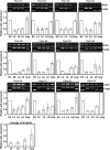
Infection course analysis of the stability of the pds gene fragment and pds mRNA silencing following biolistic or rub inoculation of FoMV-PDS in sweet corn plants (Golden × Bantam). The gel images show RT-PCR analyses for the pds insert stability in FoMV-PDS-infected plants. The top gel image is the RT-PCR control, showing amplification of a single maize actin mRNA fragment in all samples; the bottom gel image shows RT-PCR amplification across the FoMV cloning site. EV indicates the FoMV-V empty vector that carries no insert; L4, L6, L9, and Ltop indicate the leaf number that the sample was taken from. The bar graphs display the relative expression levels of pds mRNA in the indicated leaves determined by real-time qRT-PCR. B1 to B9 indicate independent plants that were biolistically inoculated, and R1 to R3 indicate independent plants that were rub inoculated. The graph at bottom left shows the relative expression of pds in each leaf sample type averaged over all plants. Error bars indicate the sd of three technical replicates for each individual plant, except for in the graph at bottom left, in which they indicate the sd for all plants. Error bars are not indicated for the EV samples, because the expression level of each FoMV-V sample was divided by itself to obtain 1.
Figure 6.
Figure 6.
Silencing les22 using the FoMV VIGS vector in sweet corn (Golden × Bantam). A, Leaf from a representative plant infected with the FoMV vector carrying a 330-bp fragment of les22. Bar = 1 cm. B, The top images show closeup views of sections of plants mock inoculated (Mock) or infected with FoMV-V empty vector or FoMV-Les22. The bottom images show the leaves after staining with Trypan Blue. Bar = 0.5 cm.
Figure 7.
Figure 7.
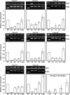
Infection course analysis of the stability of the les22 gene fragment and les22 mRNA silencing following biolistic or rub inoculation of FoMV-Les22 in sweet corn plants (Golden × Bantam). The gel images show RT-PCR analyses for the les22 insert stability in FoMV-Les22-infected plants. The top gel image is the RT-PCR control, showing amplification of a single maize actin mRNA fragment in all samples; the bottom gel image shows RT-PCR amplification across the FoMV cloning site. EV indicates the FoMV-V empty vector that carries no insert; L4, L6, L9, and Ltop indicate the leaf number that the sample was taken from. The bar graphs display the relative expression level of les22 mRNA in the indicated leaves determined by real-time qRT-PCR. B1 to B6 indicate independent plants that were biolistically inoculated, and R1 and R2 indicate independent plants that were rub inoculated. The graph at bottom right shows the relative expression of les22 in each leaf sample type averaged over all plants. Error bars indicate the sd of three technical replicates for each individual plant, except for in the graph the bottom right, in which they indicate the sd for all plants. Error bars are not indicated for the EV samples, because the expression level of each FoMV-V sample was divided by itself to obtain 1.
Figure 8.
Figure 8.
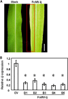
VIGS of ij using the FoMV vector. A, Sweet corn (Golden × Bantam) plants were mock inoculated (left) or biolistically inoculated with pFoMV-Ij, which carries a 231-bp fragment of maize ij (right). The leaf at right (leaf 6) shows white stripes caused by ij silencing. The white stripe at the leaf margin is highlighted in the red box. Bar = 1 cm. B, Real-time qRT-PCR analysis of ij expression in FoMV-V empty vector (EV) and FoMV-Ij-infected sweet corn (Golden × Bantam) plants. Significant suppression of ij mRNA transcripts is detected in systemic leaves of plants that were biolistically inoculated with pFoMV-Ij (*, P < 0.05 compared with the empty vector by Student’s t test). B1 to B5 indicate the sixth leaf of five different biolistically inoculated plants). Error bars indicate the SD of three technical replicates for each individual sample.
Figure 9.
Figure 9.
FoMV infection of maize inbred lines, sorghum, and green foxtail. A, Mosaic symptoms caused by FoMV-V infection were observed on systemic leaves of maize inbred lines (B73, B101, W22CC, K55, FR1064, B104, A188, and W64A), sorghum, and green foxtail but not on the maize inbred lines Mo47 or Mo17. Bars = 1 cm. B, RT-PCR amplification of a FoMV-specific PCR product confirmed FoMV infection in maize inbred lines (B73, B101, W22CC, K55, FR1064, B104, A188, W64A, and Mo47), sorghum, and green foxtail but not in the maize inbred line Mo17 (a sweet corn sample infected with FoMV is included as a positive control). The FoMV genomic fragment can be detected only in plants inoculated with FoMV-V but not in mock-treated plants. The actin gene from maize, sorghum, or green foxtail was included as an internal control.

Similar articles

Cited by

References

    1. Avesani L, Marconi G, Morandini F, Albertini E, Bruschetta M, Bortesi L, Pezzotti M, Porceddu A (2007) Stability of Potato virus X expression vectors is related to insert size: implications for replication models and risk assessment. Transgenic Res 16: 587–597 - PubMed
    1. Bancroft JB, Rouleau M, Johnston R, Prins L, Mackie GA (1991) The entire nucleotide sequence of foxtail mosaic virus RNA. J Gen Virol 72: 2173–2181 - PubMed
    1. Barrière Y, Argillier O (1993) Brown-midrib genes of maize: a review. Agronomie 13: 865–876
    1. Becker A. (2013) Virus-induced gene silencing: methods and protocols. Methods Mol Biol 975: 221
    1. Benavente LM, Ding XS, Redinbaugh MG, Nelson RS, Balint-Kurti P (2012) Virus-induced gene silencing in diverse maize lines using the Brome mosaic virus-based silencing vector. Maydica 57: 206–214

Publication types