

Requirement for basement membrane laminin α5 during urethral and external genital development
- PMID: 27208857
- PMCID: PMC4995130
- DOI: 10.1016/j.mod.2016.05.004
Requirement for basement membrane laminin α5 during urethral and external genital development
Abstract
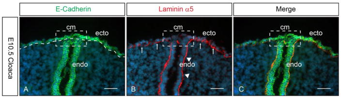
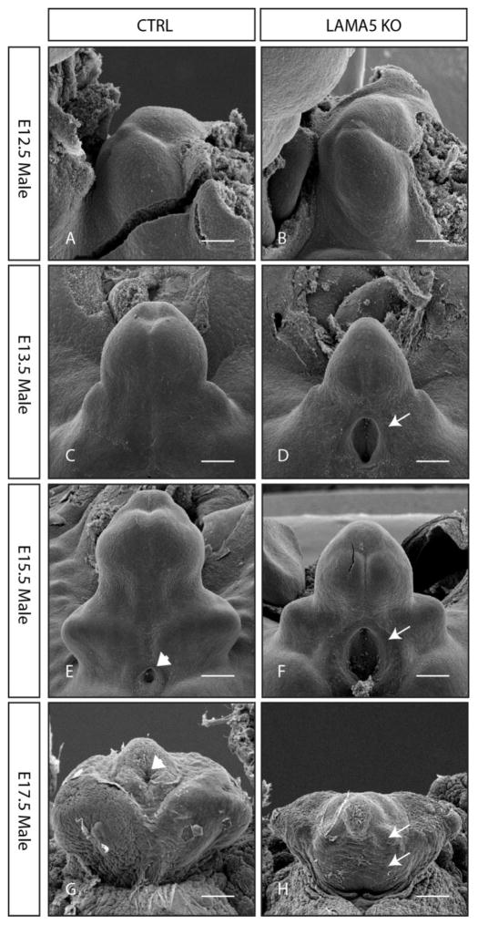
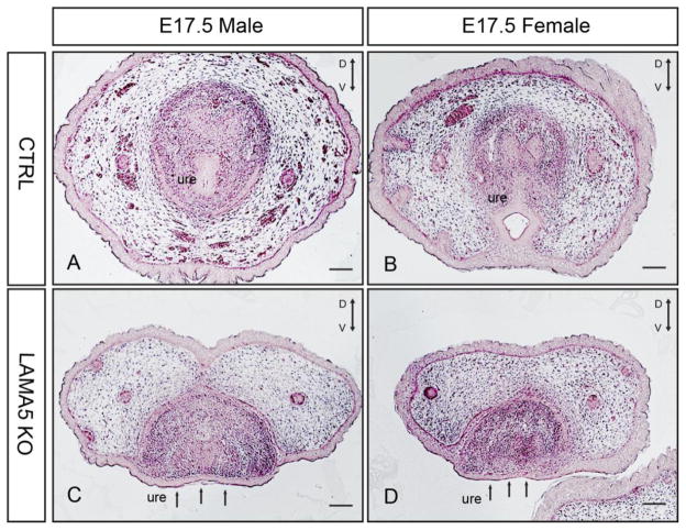
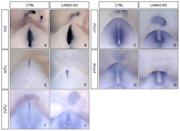
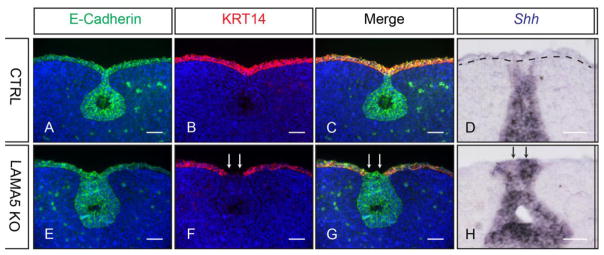
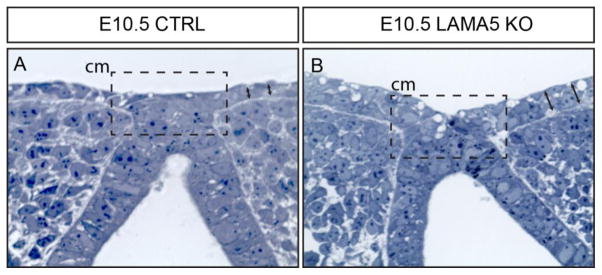
Hypospadias, a congenital malformation of the penis characteristic of an abnormal urethral orifice, affects 1 in every 125 boys, and its incidence is rising. Herein we test the hypothesis that the basement membrane protein laminin α5 (LAMA5) plays a key role in the development of the mouse genital tubercle, the embryonic anlage of the external genitalia. Using standard histological analyses and electron microscopy, we characterized the morphology of the external genitalia in Lama5 knockout (LAMA5-KO) mouse embryos during both androgen-independent genital tubercle development and androgen-mediated sexual differentiation. We compared regulatory gene expression between control and LAMA5-KO by in situ hybridization. We also examined the epithelial structure of the mutant genital tubercle using immunofluorescence staining and histological analyses of semi-thin sections. We found that Lama5 was expressed in both ectodermal and endodermal epithelia of the cloaca. The LAMA5-KO displayed a profound external genital malformation in which the genital tubercle was underdeveloped with a large ectopic orifice at the proximal end. In older embryos, the urethra failed to form a tubular structure and was left completely exposed. These defects were not associated with a significant alteration in regulatory gene expression, but rather with a defective ectodermal epithelium and an abnormal disintegration of the cloacal membrane. We conclude that LAMA5 is required in the basement membrane to maintain normal architecture of the ventral ectoderm during genital tubercle development, which is essential for the formation of a tubular urethra. Perturbation of LAMA5, and possibly other basement membrane components, may cause hypospadias in humans.
Keywords: Basement membrane; External genitalia; Hypospadias; Laminin α5; Urethra.
Copyright © 2016 Elsevier B.V. All rights reserved.
Figures
Similar articles
-
Multiphasic and tissue-specific roles of sonic hedgehog in cloacal septation and external genitalia development.Development. 2009 Dec;136(23):3949-57. doi: 10.1242/dev.042291. Development. 2009. PMID: 19906862 Free PMC article.
-
Tissue-specific roles of Fgfr2 in development of the external genitalia.Development. 2015 Jun 15;142(12):2203-12. doi: 10.1242/dev.119891. Development. 2015. PMID: 26081573 Free PMC article.
-
Sexually dimorphic expression of Mafb regulates masculinization of the embryonic urethral formation.Proc Natl Acad Sci U S A. 2014 Nov 18;111(46):16407-12. doi: 10.1073/pnas.1413273111. Epub 2014 Oct 31. Proc Natl Acad Sci U S A. 2014. PMID: 25362053 Free PMC article.
-
Developmental mutant mouse models for external genitalia formation.Congenit Anom (Kyoto). 2019 May;59(3):74-80. doi: 10.1111/cga.12319. Epub 2018 Dec 16. Congenit Anom (Kyoto). 2019. PMID: 30554442 Review.
-
Regulatory roles of epithelial-mesenchymal interaction (EMI) during early and androgen dependent external genitalia development.Differentiation. 2019 Nov-Dec;110:29-35. doi: 10.1016/j.diff.2019.08.004. Epub 2019 Sep 11. Differentiation. 2019. PMID: 31590136 Review.
Cited by
-
Spatiotemporal map of key signaling factors during early penis development.Dev Dyn. 2022 Apr;251(4):609-624. doi: 10.1002/dvdy.433. Epub 2021 Nov 6. Dev Dyn. 2022. PMID: 34697862 Free PMC article. Review.
-
Identification of Small Regions of Overlap from Copy Number Variable Regions in Patients with Hypospadias.Int J Mol Sci. 2022 Apr 12;23(8):4246. doi: 10.3390/ijms23084246. Int J Mol Sci. 2022. PMID: 35457073 Free PMC article.
-
Temporal, spatial, and genetic regulation of external genitalia development.Differentiation. 2019 Nov-Dec;110:1-7. doi: 10.1016/j.diff.2019.08.003. Epub 2019 Sep 10. Differentiation. 2019. PMID: 31521888 Free PMC article. Review.
-
Hedgehog Signaling for Urogenital Organogenesis and Prostate Cancer: An Implication for the Epithelial-Mesenchyme Interaction (EMI).Int J Mol Sci. 2019 Dec 20;21(1):58. doi: 10.3390/ijms21010058. Int J Mol Sci. 2019. PMID: 31861793 Free PMC article. Review.
-
Etiology of Hypospadias: A Comparative Review of Genetic Factors and Developmental Processes Between Human and Animal Models.Res Rep Urol. 2020 Dec 24;12:673-686. doi: 10.2147/RRU.S276141. eCollection 2020. Res Rep Urol. 2020. PMID: 33381468 Free PMC article. Review.
References
-
- Baskin LS, Erol A, Jegatheesan P, Li Y, Liu W, Cunha GR. Urethral seam formation and hypospadias. Cell and tissue research. 2001;305:379–387. - PubMed
-
- Ettner N, Gohring W, Sasaki T, Mann K, Timpl R. The N-terminal globular domain of the laminin alpha1 chain binds to alpha1beta1 and alpha2beta1 integrins and to the heparan sulfate-containing domains of perlecan. FEBS Lett. 1998;430:217–221. - PubMed
-
- Gatti JM, Kirsch AJ, Troyer WA, Perez-Brayfield MR, Smith EA, Scherz HC. Increased incidence of hypospadias in small-for-gestational age infants in a neonatal intensive-care unit. BJU international. 2001;87:548–550. - PubMed
-
- Haraguchi R, Mo R, Hui C, Motoyama J, Makino S, Shiroishi T, Gaffield W, Yamada G. Unique functions of Sonic hedgehog signaling during external genitalia development. Development. 2001;128:4241–4250. - PubMed
Publication types
MeSH terms
Substances
Grants and funding
LinkOut - more resources
Full Text Sources
Other Literature Sources
Molecular Biology Databases
Research Materials
