

Monosodium urate crystals induce oxidative stress in human synoviocytes
- PMID: 27209322
- PMCID: PMC4875700
- DOI: 10.1186/s13075-016-1012-3
Monosodium urate crystals induce oxidative stress in human synoviocytes
Abstract
Background: Gout is the most common inflammatory arthropathy of metabolic origin and it is characterized by intense inflammation, the underlying mechanisms of which are unknown. The aim of this study was to evaluate the oxidative stress in human fibroblast-like synoviocytes (FLS) exposed to monosodium urate (MSU) crystals, which trigger an inflammatory process.
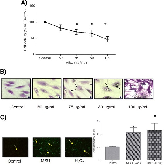
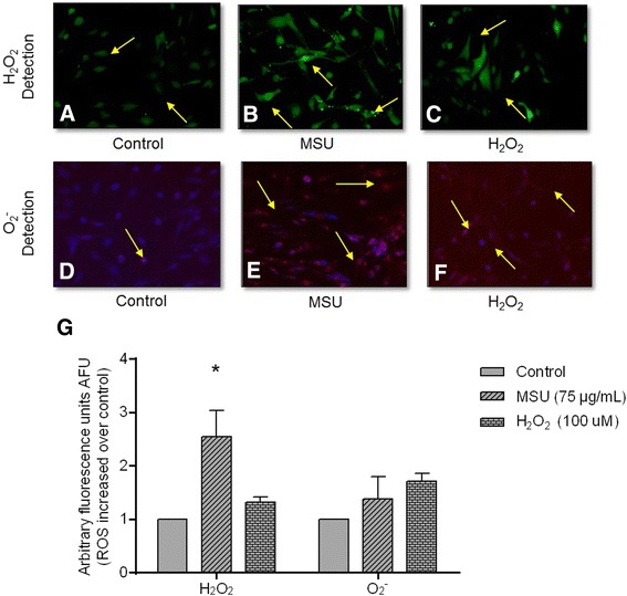
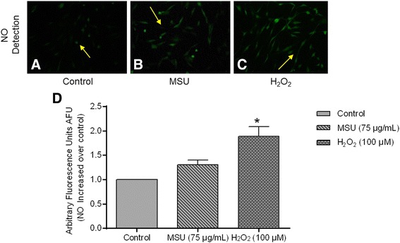
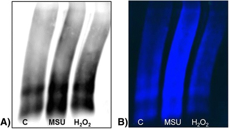
Methods: Human FLS isolated from synovial tissue explants were stimulated with MSU crystals (75 μg/mL) for 24 h. Cellular viability was evaluated by crystal violet staining, apoptosis was assessed using Annexin V, and the cellular content of reactive oxygen species (ROS) and nitrogen species (RNS) (O2 (-), H2O2, NO) was assessed with image-based cytometry and fluorometric methods. In order to determine protein oxidation levels, protein carbonyls were detected through oxyblot analysis, and cell ultrastructural changes were assessed by transmission electron microscopy.
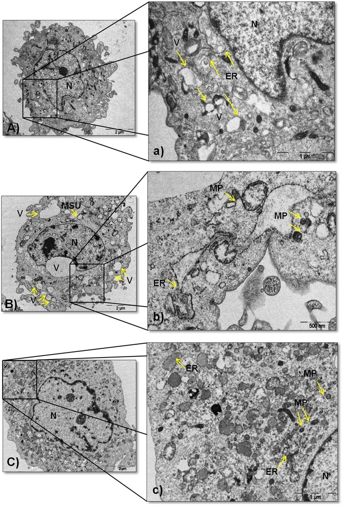
Results: The viability of FLS exposed to MSU crystals decreased by 30 % (P < 0.05), while apoptosis increased by 42 % (P = 0.01). FLS stimulated with MSU crystals exhibited a 2.1-fold increase in H2O2 content and a 1.5-fold increase in O2 (-) and NO levels. Oxyblots revealed that the spots obtained from FLS protein lysates exposed to MSU crystals exhibited protein carbonyl immunoreactivity, which reflects the presence of oxidatively modified proteins. Concomitantly, MSU crystals triggered the induction of changes in the morphostructure of FLS, such as the thickening and discontinuity of the endoplasmic reticulum, and the formation of vacuoles and misfolded glycoproteins.
Conclusions: Our results prove that MSU crystals induce the release of ROS and RNS in FLS, subsequently oxidizing proteins and altering the cellular oxidative state of the endoplasmic reticulum, which results in FLS apoptosis.
Keywords: Gout; Monosodium urate crystals; Oxidative stress; Synoviocytes.
Figures
Similar articles
-
High-density lipoproteins downregulate CCL2 production in human fibroblast-like synoviocytes stimulated by urate crystals.Arthritis Res Ther. 2010;12(1):R23. doi: 10.1186/ar2930. Epub 2010 Feb 11. Arthritis Res Ther. 2010. PMID: 20149224 Free PMC article.
-
Role of the NLRP3 inflammasome in the transient release of IL-1β induced by monosodium urate crystals in human fibroblast-like synoviocytes.J Inflamm (Lond). 2015 Apr 10;12:30. doi: 10.1186/s12950-015-0070-7. eCollection 2015. J Inflamm (Lond). 2015. PMID: 25897296 Free PMC article.
-
Monosodium urate crystals reduce osteocyte viability and indirectly promote a shift in osteocyte function towards a proinflammatory and proresorptive state.Arthritis Res Ther. 2018 Sep 10;20(1):208. doi: 10.1186/s13075-018-1704-y. Arthritis Res Ther. 2018. PMID: 30201038 Free PMC article.
-
Molecular basis of oxidative stress in gouty arthropathy.Clin Rheumatol. 2015 Oct;34(10):1667-72. doi: 10.1007/s10067-015-2933-y. Epub 2015 Apr 9. Clin Rheumatol. 2015. PMID: 25854697 Review.
-
Gout: epitome of painful arthritis.Metabolism. 2010 Oct;59 Suppl 1:S32-6. doi: 10.1016/j.metabol.2010.07.009. Metabolism. 2010. PMID: 20837191 Review.
Cited by
-
TLR4 sensing of IsdB of Staphylococcus aureus induces a proinflammatory cytokine response via the NLRP3-caspase-1 inflammasome cascade.mBio. 2024 Jan 16;15(1):e0022523. doi: 10.1128/mbio.00225-23. Epub 2023 Dec 19. mBio. 2024. PMID: 38112465 Free PMC article.
-
Gout and the Risk of Incident Obstructive Sleep Apnea in Adults 65 Years or Older: An Observational Study.J Clin Sleep Med. 2018 Sep 15;14(9):1521-1527. doi: 10.5664/jcsm.7328. J Clin Sleep Med. 2018. PMID: 30176977 Free PMC article.
-
CXCL5 activates CXCR2 in nociceptive sensory neurons to drive joint pain and inflammation in experimental gouty arthritis.Nat Commun. 2024 Apr 16;15(1):3263. doi: 10.1038/s41467-024-47640-7. Nat Commun. 2024. PMID: 38627393 Free PMC article.
-
Prooxidant effect of uric acid on human leukocytic DNA: An in vitro and ex vivo study.Turk J Biol. 2025 Jan 2;49(2):175-184. doi: 10.55730/1300-0152.2735. eCollection 2025. Turk J Biol. 2025. PMID: 40365102 Free PMC article.
-
Neutrophil Extracellular Traps and By-Products Play a Key Role in COVID-19: Pathogenesis, Risk Factors, and Therapy.J Clin Med. 2020 Sep 11;9(9):2942. doi: 10.3390/jcm9092942. J Clin Med. 2020. PMID: 32933031 Free PMC article. Review.
References
MeSH terms
Substances
LinkOut - more resources
Full Text Sources
Other Literature Sources
Medical
