Review
. 2016 Aug;60(2):65-73.
doi: 10.1016/j.ceca.2016.05.001. Epub 2016 May 4.

Subcellular ROS imaging methods: Relevance for the study of calcium signaling

Affiliations
Review

Subcellular ROS imaging methods: Relevance for the study of calcium signaling

David M Booth et al. Cell Calcium. 2016 Aug.

Abstract

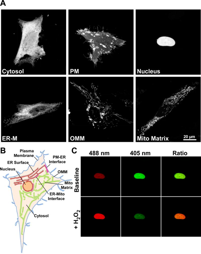
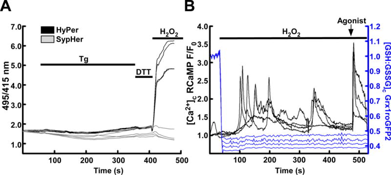
Recent advances in genetically encoded fluorescent probes have dramatically increased the toolkit available for imaging the intracellular environment. Perhaps the biggest improvements have been made in sensing specific reactive oxygen species (ROS) and redox changes under physiological conditions. The new generation of probes may be targeted to a wide range of subcellular environments. By targeting such probes to compartments and organelle surfaces they may be exposed to environments, which support local signal transduction and regulation. The close apposition of the endoplasmic reticulum (ER) with mitochondria and other organelles forms such a local environment where Ca(2+) dynamics are greatly enhanced compared to the bulk cytosol. We describe here how newly developed genetically encoded redox indicators (GERIs) might be used to monitor ROS and probe their interaction with Ca(2+) at both global and local level.

Keywords: Grx1-roGFP2; H(2)O(2); HyPer; Redox; SypHer.


Figures

Figure 1
Figure 1
The genetically encoded redox indicator HyPer is a functional H2O2 sensor when targeted to diverse subcellular environments. (A) Confocal images of HeLa cells transfected with HyPer targeted to the cytosol, cytosolic surface of the plasma membrane (PM), nucleus, cytosolic surface of the endoplasmic reticulum membrane (ER-M), cytosolic surface of the outer mitochondrial membrane (OMM) and mitochondrial matrix. Images are the sum of 488 & 405 nm excitation with a 1 pixel median filter step. (B) Scheme depicting some subcellular organelles viable for probe targeting, Plasma membrane (blue), ER membrane (red), Nucleus (orange), outer mitochondrial membrane (OMM, green), mitochondrial matrix (light green) and cytosol (tan). Close appositions of the ER with the plasma membrane (PM-ER interface), mitochondria (ER-mito interface) are highlighted (red). (C) HeLa cell expressing HyPer targeted to the nucleus. Fluorescence intensity images of 488 nm excitation (red) and 405 nm excitation (green) before and after addition of H2O2 (100 μM). Overlaid images (right) show increase in 488 nm fluorescence (red) and decrease in 405 nm fluorescence (green) following H2O2 addition.
Figure 2
Figure 2
Calcium and redox status may be imaged simultaneously. (A) HEK293 cells transiently transfected with HyPer (black) or the redox-desensitized derivative SypHer (grey) treated with the sarcoendoplasmic reticulum Ca2+ ATPase inhibitor thapsigargin (Tg, 2μM) to induce cytosolic and mitochondrial Ca2+ elevations. Small shifts in HyPer and SypHer occur in response to Tg, but a lack of response to DTT suggests that any changes are pH derived. H2O2 (200 μM) is added at the end of the experiment to demonstrate maximal response. (B) HEK293 cells transiently transfected with Grx1roGFP2 (blue) and RCaMP (black) targeted to the cytosol. Addition of H2O2 (200 μM) oxidizes the GSH:GSSG ratio, subsequently, dysregulated H2O2-induced cytosolic Ca2+ elevations occur. An IP3-linked agonist, carbachol (CCh, 100 μM) is added at the end of the experiment to demonstrate the effect of maximal IP3 receptor-mediated Ca2+ release.

References

    1. Berridge MJ, Lipp P, Bootman MD. The versatility and universality of calcium signalling. Nat Rev Mol Cell Biol. 2000;1(1):11–21. - PubMed
    1. Berridge MJ, Galione A. Cytosolic calcium oscillators. FASEB J. 1988;2(15):3074–82. - PubMed
    1. Putney JW, Jr, Thomas AP. Calcium signaling: double duty for calcium at the mitochondrial uniporter. Curr Biol. 2006;16(18):R812–5. - PMC - PubMed
    1. Tinel H, et al. Active mitochondria surrounding the pancreatic acinar granule region prevent spreading of inositol trisphosphate-evoked local cytosolic Ca(2+) signals. EMBO J. 1999;18(18):4999–5008. - PMC - PubMed
    1. Parker I, Ivorra I. Localized all-or-none calcium liberation by inositol trisphosphate. Science. 1990;250(4983):977–9. - PubMed

Publication types

Substances