. 2016 May 23:6:26521.
doi: 10.1038/srep26521.

Proteomic profiling of small-molecule inhibitors reveals dispensability of MTH1 for cancer cell survival

Affiliations

Proteomic profiling of small-molecule inhibitors reveals dispensability of MTH1 for cancer cell survival

Tatsuro Kawamura et al. Sci Rep. .

Abstract

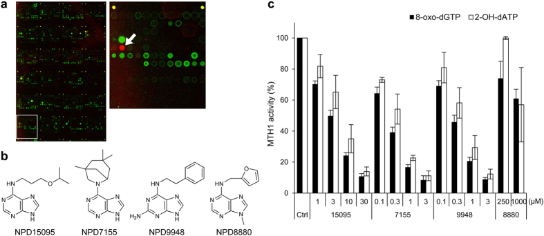
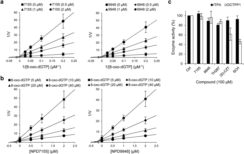
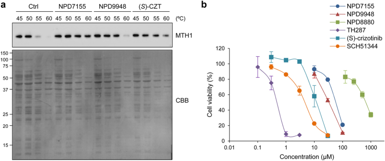
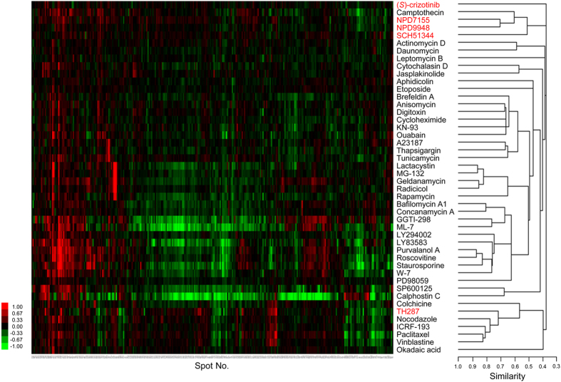
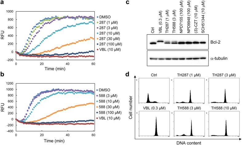
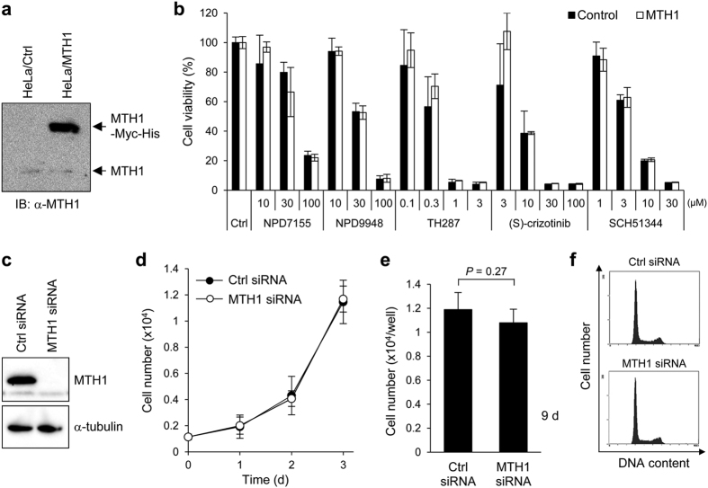
Since recent publications suggested that the survival of cancer cells depends on MTH1 to avoid incorporation of oxidized nucleotides into the cellular DNA, MTH1 has attracted attention as a potential cancer therapeutic target. In this study, we identified new purine-based MTH1 inhibitors by chemical array screening. However, although the MTH1 inhibitors identified in this study targeted cellular MTH1, they exhibited only weak cytotoxicity against cancer cells compared to recently reported first-in-class inhibitors. We performed proteomic profiling to investigate the modes of action by which chemically distinct MTH1 inhibitors induce cancer cell death, and found mechanistic differences among the first-in-class MTH1 inhibitors. In particular, we identified tubulin as the primary target of TH287 and TH588 responsible for the antitumor effects despite the nanomolar MTH1-inhibitory activity in vitro. Furthermore, overexpression of MTH1 did not rescue cells from MTH1 inhibitor-induced cell death, and siRNA-mediated knockdown of MTH1 did not suppress cancer cell growth. Taken together, we conclude that the cytotoxicity of MTH1 inhibitors is attributable to off-target effects and that MTH1 is not essential for cancer cell survival.


Figures

Figure 1
Figure 1. Identification of purine-based MTH1 inhibitors by chemical array screening.
(a) Representative fluorescent image of the chemical arrays (left) and magnified image of the area in a white square (right). Spot of NPD15095 is indicated by a white arrow. (b) Chemical structures of purine-based MTH1 inhibitors. (c) Effects of NPD15095 (15095), NPD7155 (7155), NPD9948 (9948), and NPD8880 (8880) on the catalytic activity of MTH1. 8-oxo-dGTP and 2-OH-dATP were used as substrates. Data are shown as mean ± s.d. from three independent experiments.
Figure 2
Figure 2. Kinetics and selectivity of NPD7155 and NPD9948.
(a,b) Kinetic analysis of NPD7155 (7155) and NPD9948 (9948) against MTH1. Lineweaver–Burk plot of reciprocal of initial velocity vs. reciprocal of varying 8-oxo-dGTP concentrations (a) and Dixon plot of reciprocal of initial velocity vs. varying concentrations of NPD7155 or NPD9948 (b). Data are shown as mean ± s.e.m. from three independent experiments. (c) Effects of NPD7155, NPD9948, TH287, (S)-crizotinib [(S)-CZT], and SCH51344 (SCH) on the catalytic activities of ITPA and DCTPP1 in vitro. Data are shown as mean ± s.d. from three independent experiments.
Figure 3
Figure 3. NPD7155 and NPD9948 exhibit less potent cytotoxicity.
(a) Target engagement of NPD7155 and NPD9948 to MTH1 protein in intact HeLa cells. HeLa cells were treated with NPD7155 (300 μM), NPD9948 (300 μM), or (S)-crizotinib (30 μM) for 1 h, collected, heated at the indicated temperatures, and lysed. Soluble proteins collected in the supernatant were subjected to western blot analysis to detect MTH1, followed by staining with Coomassie brilliant blue (CBB). (b) Viability of HeLa cells treated with MTH1 inhibitors for 72 h. Data are shown as mean ± s.d.
Figure 4
Figure 4. Proteomic profiling shows mechanistic differences among MTH1 inhibitors.
HeLa cells were treated with NPD7155 (300 μM), NPD9948 (300 μM), SCH51344 (60 μM), (S)-crizotinib (20 μM), or TH287 (3 μM) for 18 h. Proteomic analysis of cell lysates was performed by the 2-D DIGE system. Quantitative data of the common 296 spots (x-axis) derived from MTH1 inhibitors and those of 41 well-characterized compounds were analyzed by hierarchical clustering. As shown in the scale bar for the heat map, intensities of red and green coloration indicate an increased or decreased log-fold (natural base) normalized volume, respectively, of spots for each compound. The vertical axis represents compounds. The horizontal axes of the heat map and dendrogram represent spot number and cosine similarity between clusters, respectively.
Figure 5
Figure 5. TH287 and TH588 inhibit tubulin polymerization.
(a,b) Inhibitory effects of TH287 (a) and TH588 (b) on tubulin polymerization in vitro. Vinblastine (VBL) was used as a positive control. (c) Detection of phosphorylated Bcl-2 in HeLa cells treated with compounds for 24 h. (d) Cell cycle analysis of HeLa cells treated with compounds for 24 h.
Figure 6
Figure 6. MTH1 inhibition does not affect cancer cell growth.
(a,b) Overexpression of MTH1 did not rescue cells from MTH1 inhibitors-induced cell death. HeLa cells transiently overexpressing human MTH1 and the control counterparts were treated with MTH1 inhibitors for 84 h. The expression levels of MTH1 proteins in both cells were analyzed by western blot (a). The cell viability was examined by WST-8 assay. Data are shown as mean ± s.d. There was no statistically significant difference between the viability of MTH1-overexpressing HeLa cells and that of the control counterparts following treatment with MTH1 inhibitors at all concentrations tested (ANOVA followed by Games-Howell test) (b). (cf) Knockdown of MTH1 did not affect cell survival. HeLa cells were transfected with 20 nM siRNA for 72 h, and the knockdown of MTH1 was analyzed by western blot (c). Cell growth of HeLa cells transfected with 20 nM siRNA for the indicated times (d,e). Data are shown as mean ± s.d. Statistical analysis was performed using Student’s t-test. Cell cycle analysis of HeLa cells transfected with 20 nM siRNA for 96 h (f).

Similar articles

  • Validation and development of MTH1 inhibitors for treatment of cancer.
    Warpman Berglund U, Sanjiv K, Gad H, Kalderén C, Koolmeister T, Pham T, Gokturk C, Jafari R, Maddalo G, Seashore-Ludlow B, Chernobrovkin A, Manoilov A, Pateras IS, Rasti A, Jemth AS, Almlöf I, Loseva O, Visnes T, Einarsdottir BO, Gaugaz FZ, Saleh A, Platzack B, Wallner OA, Vallin KS, Henriksson M, Wakchaure P, Borhade S, Herr P, Kallberg Y, Baranczewski P, Homan EJ, Wiita E, Nagpal V, Meijer T, Schipper N, Rudd SG, Bräutigam L, Lindqvist A, Filppula A, Lee TC, Artursson P, Nilsson JA, Gorgoulis VG, Lehtiö J, Zubarev RA, Scobie M, Helleday T. Warpman Berglund U, et al. Ann Oncol. 2016 Dec;27(12):2275-2283. doi: 10.1093/annonc/mdw429. Epub 2016 Nov 8. Ann Oncol. 2016. PMID: 27827301
  • MTH1 inhibition eradicates cancer by preventing sanitation of the dNTP pool.
    Gad H, Koolmeister T, Jemth AS, Eshtad S, Jacques SA, Ström CE, Svensson LM, Schultz N, Lundbäck T, Einarsdottir BO, Saleh A, Göktürk C, Baranczewski P, Svensson R, Berntsson RP, Gustafsson R, Strömberg K, Sanjiv K, Jacques-Cordonnier MC, Desroses M, Gustavsson AL, Olofsson R, Johansson F, Homan EJ, Loseva O, Bräutigam L, Johansson L, Höglund A, Hagenkort A, Pham T, Altun M, Gaugaz FZ, Vikingsson S, Evers B, Henriksson M, Vallin KS, Wallner OA, Hammarström LG, Wiita E, Almlöf I, Kalderén C, Axelsson H, Djureinovic T, Puigvert JC, Häggblad M, Jeppsson F, Martens U, Lundin C, Lundgren B, Granelli I, Jensen AJ, Artursson P, Nilsson JA, Stenmark P, Scobie M, Berglund UW, Helleday T. Gad H, et al. Nature. 2014 Apr 10;508(7495):215-21. doi: 10.1038/nature13181. Epub 2014 Apr 2. Nature. 2014. PMID: 24695224
  • MTH1 deficiency selectively increases non-cytotoxic oxidative DNA damage in lung cancer cells: more bad news than good?
    Abbas HHK, Alhamoudi KMH, Evans MD, Jones GDD, Foster SS. Abbas HHK, et al. BMC Cancer. 2018 Apr 16;18(1):423. doi: 10.1186/s12885-018-4332-7. BMC Cancer. 2018. PMID: 29661172 Free PMC article.
  • Role of MTH1 in oxidative stress and therapeutic targeting of cancer.
    Taiyab A, Ashraf A, Sulaimani MN, Rathi A, Shamsi A, Hassan MI. Taiyab A, et al. Redox Biol. 2024 Nov;77:103394. doi: 10.1016/j.redox.2024.103394. Epub 2024 Oct 11. Redox Biol. 2024. PMID: 39418911 Free PMC article. Review.
  • Clinical Significance of NUDT1 (MTH1) Across Cancer Types.
    Misiak R, Białkowski K, Dondajewska E. Misiak R, et al. Int J Mol Sci. 2025 May 27;26(11):5137. doi: 10.3390/ijms26115137. Int J Mol Sci. 2025. PMID: 40507948 Free PMC article. Review.

Cited by

References

    1. Trachootham D., Alexandre J. & Huang P. Targeting cancer cells by ROS-mediated mechanisms: a radical therapeutic approach? Nat. Rev. Drug Discov. 8, 579–591 (2009). - PubMed
    1. Hu Y. et al. Mitochondrial manganese-superoxide dismutase expression in ovarian cancer: role in cell proliferation and response to oxidative stress. J. Biol. Chem. 280, 39485–39492 (2005). - PubMed
    1. Xia C. et al. Reactive oxygen species regulate angiogenesis and tumor growth through vascular endothelial growth factor. Cancer Res. 67, 10823–10830 (2007). - PubMed
    1. Luo M., He H., Kelley M. R. & Georgiadis M. M. Redox regulation of DNA repair: implications for human health and cancer therapeutic development. Antioxid. Redox Signal 12, 1247–1269 (2010). - PMC - PubMed
    1. Topal M. D. & Baker M. S. DNA precursor pool: a significant target for N-methyl-N-nitrosourea in C3H/10T1/2 clone 8 cells. Proc. Natl Acad. Sci. USA 79, 2211–2215 (1982). - PMC - PubMed

Publication types

MeSH terms