. 2016 Jul;49(1):153-63.
doi: 10.3892/ijo.2016.3525. Epub 2016 May 16.

Overcoming resistance to TRAIL-induced apoptosis in solid tumor cells by simultaneously targeting death receptors, c-FLIP and IAPs

Affiliations

Overcoming resistance to TRAIL-induced apoptosis in solid tumor cells by simultaneously targeting death receptors, c-FLIP and IAPs

Ying Huang et al. Int J Oncol. 2016 Jul.

Abstract

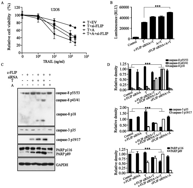
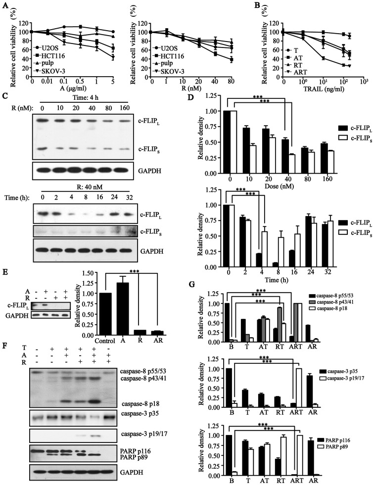
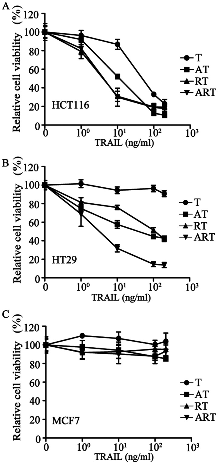
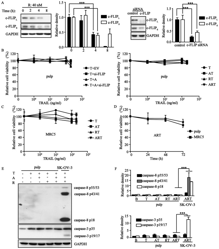
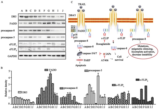
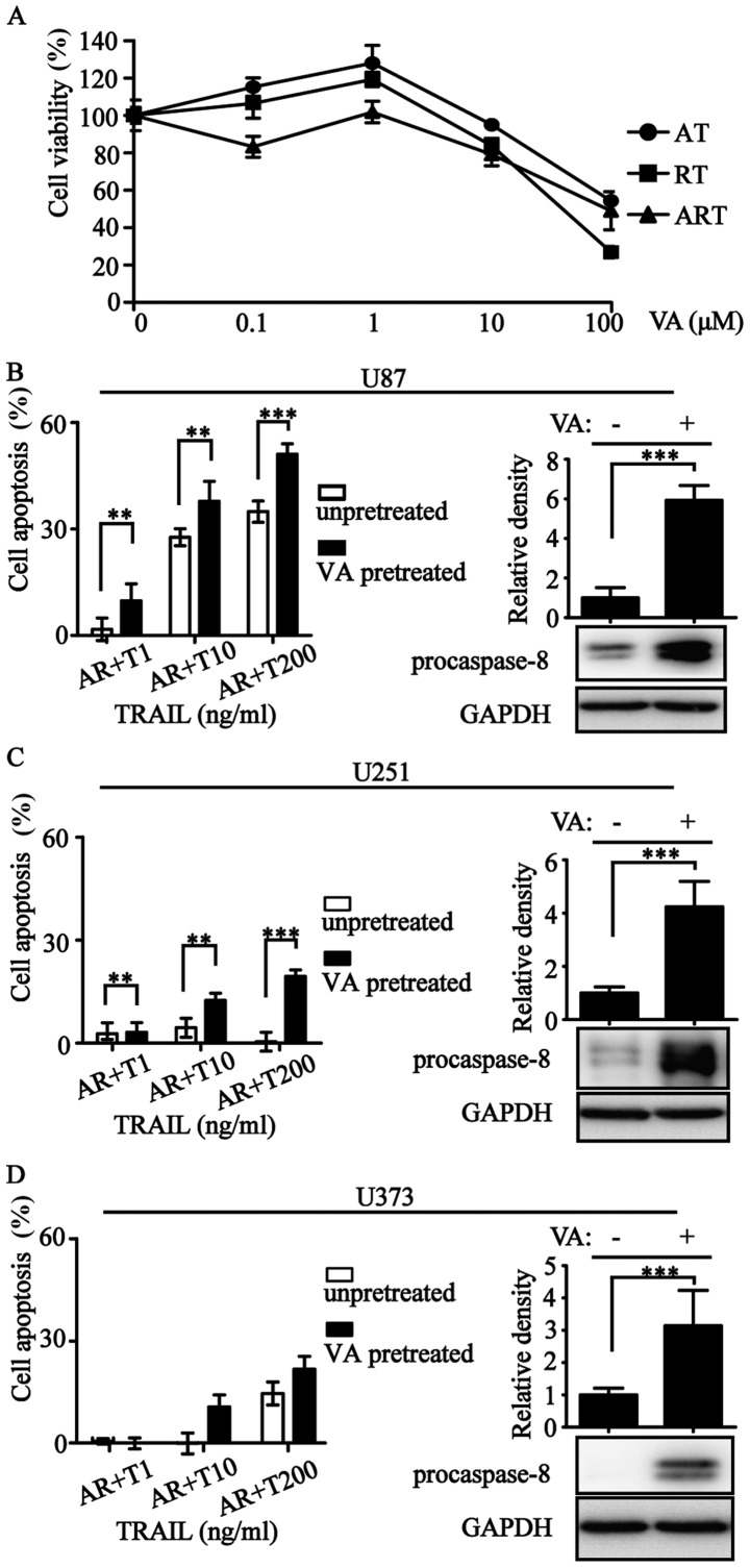
The discovery of the TRAIL protein and its death receptors DR4/5 changed the horizon of cancer research because TRAIL specifically kills cancer cells. However, the validity of TRAIL-based cancer therapies has yet to be established, as most cancer cells are TRAIL-resistant. In this report, we demonstrate that TRAIL-resistance of many cancer cell lines can be overcome after siRNA- or rocaglamide-mediated downregulation of c-FLIP expression and simultaneous inhibition of IAPs activity using AT406, a pan-antagonist of IAPs. Combined triple actions of the TRAIL, the IAPs inhibitor, AT406, and the c-FLIP expression inhibitor, rocaglamide (ART), markedly improve TRAIL-induced apoptotic effects in most solid cancer cell lines through the activation of an extrinsic apoptosis pathway. Furthermore, this ART combination does not harm normal cells. Among the 18 TRAIL-resistant cancer cell lines used, 15 cell lines become sensitive or highly sensitive to ART, and two out of three glioma cell lines exhibit high resistance to ART treatment due to very low levels of procaspase-8. This study provides a rationale for the development of TRAIL-induced apoptosis-based cancer therapies.


Figures

Figure 1
Figure 1
The combination of AT406 and downregulation of c-FLIPL/S by c-FLIP-siRNA sensitizes the osteosarcoma cells U2OS to TRAIL-induced apoptosis. (A) The osteosarcoma cells U2OS were treated with different concentrations of TRAIL for 24 h. In three other concentration-response experiments, cells were pre-treated with AT406 (A) for 1 h and with or without transient transfection with c-FLIP-siRNA for 24 h. The cell viabilities were determined by an MTT assay. The results are shown as the mean ± SEM of three separate experiments. (B) Caspase activity of U2OS cells in response to TRAIL with or without pre-treatments of A and c-FLIP-siRNA as indicated in the graph was measured by a Caspase-Glo 3/7 assay. (C) The U2OS cells were treated with TRAIL for 5 h, and with or without pre-treatment of A for 1 h, and transient transfection with c-FLIP-siRNA for 24 h as indicated in the graph. The extrinsic apoptosis related proteins were determined by western blots and statistical analyses and are shown in (D). In all experiments if not specified: A, AT406 at 1 μg/ml; EV, control empty vector; c-FLIP-siRNA, pSuper-puro-siRNA-c-FLIP vector; T, TRAIL at 100 ng/ml. The results are shown as the means ± SEM in all bar graphs of western blots of three independent experiments (NS>0.05, ***p<0.001).
Figure 2
Figure 2
Rocaglamide behaves similarly to c-FLIP-siRNA to inhibit the expression of c-FLIPL/S and, in combination with AT406, sensitized U2OS cells for TRAIL-induced apoptosis. (A) Different cells (cancer cells and normal cells) were treated by different doses of AT406 (A) and rocaglamide (R). Cell viabilities were measured by an MTT assay. (B) Cell viabilities of U2OS in response to TRAIL with or without pre-treatments of A and R as indicated in the graph. Cell viability determined by an MTT assay in A and B are presented as the mean ± SEM of three separate experiments. (C) The osteosarcoma cells U2OS were treated with different concentrations of rocaglamide for 4 h or treated with 40 nM rocaglamide for different durations. The c-FLIPL/S expression was determined using western blots. (D) Statistical analysis of western blots in (C). (E) The U2OS cells were treated with rocaglamide and with or without AT406 for 8 h. The c-FLIPL/S expressions were determined using western blots. (F) The osteosarcoma cells U2OS were treated with TRAIL at 100 ng/ml for 5 h, and with or without pre-treatment of A at 1 μg/ml for 1 h, R at 40 nM for 4 h as indicated in the graph. The extrinsic apoptosis related proteins were determined by western blots. (G) Statistical analysis of western blots in F for caspase-3 and PARP related proteins. The bar graphs represent the mean ± SD. The results are representative of three independent experiments (NS>0.05, ***p<0.001).
Figure 3
Figure 3
Concentration-cell viability inhibition curves of TRAIL in the absence and presence of AT406 and rocaglamide, and the time course of ART triple combination treatments in cancer cell lines. (A–C) Cell viabilities of example cell lines HCT116 (A), HT29 (B) and MCF7 (C) in response to TRAIL with or without pre-treatment of A and R as indicated in the graph (A at 1 μg/ml; R at 40 nM. In each TRAIL concentration-response experiments, TRAIL treating time is 24 h). The results are shown as the mean ± SEM of three independent experiments.
Figure 4
Figure 4
Normal cell pulp and MRC5 are resistant to ART combination treatment. (A) The pulp cells were treated with 40 nM rocaglamide for different durations or with/without c-FLIP-siRNA transfection for 24 h. The c-FLIPL/S expression was determined using western blots. (B) Left, the normal cell pulp was treated with different concentrations of TRAIL for 24 h in combination with c-FLIP-siRNA (si-FLIP, transient transfection with c-FLIP-siRNA for 24 h), AT406 (A) or empty vector (EV) as indicated in the graph. (B) Right, pulp cells were treated with various concentrations of TRAIL for 24 h with or without the combination of A and R as indicated in the graph. (C) MRC5 cells were treated with various concentrations of TRAIL for 24 h with or without the combination of A and R as indicated in the graph. In experiments of B and C, cells were pretreated by R at 40 nM for 4 h, and A at 1 μg/ml for 1-h pretreatment. The cell viability was determined by an MTT assay. (D) The time course of cell viability of pulp and MRC5 cells after treatment with the ART combination. In cell viability assays, results are shown as the means ± SEM of three independent experiments. (E) The ART triple combination did not activate the extrinsic apoptosis pathway. The normal cell pulp was treated with TRAIL at 100 ng/ml for 5 h, with or without the pretreatment of A at 1 μg/ml for 1 h and R at 40 nM for 4 h as indicated in the graph. The ovarian cancer cell SK-OV-3 is shown as a positive control after ART combination treatments. (F) Statistical analysis of western blots in (E). Means ± SD are shown in all bar graphs of western blots on the right side. The results are representative of three independent experiments.
Figure 5
Figure 5
The resistance to synergistic actions of triple combination may be attributed to the low expression of procaspases. (A) TRAIL related apoptosis proteins of the 9 selected cell lines and normal cell pulp were detected by western blots without any drug treatment. (B) All of the TRAIL-related apoptosis proteins in these 9 cancer cell lines and pulp cells were compared with LOVO. (A) LOVO; (B) HCT116; (C) U2OS; (D) SK-OV-3; (E) HT29; (F) MCF7; (G) U87; (H) U251; (I) U373; (J) pulp. The results are presented in the bar graphs as the means ± SD for western blots from three independent experiments. (C) Model showing the mechanisms of sensitization of TRAIL-induced apoptosis by rocaglamide as a c-FLIP synthetic inhibitor, and AT406 as a pan-antagonist of IAPs. Abnormal expression of procaspase-8 prevents the progress of apoptosis pathway in cancer cells and cells develop resistance to both TRAIL- and ART triple combination-induced apoptosis.
Figure 6
Figure 6
The procaspase-8 expression positively correlates with TRAIL inducing apoptosis. (A) Different cells (U87, U251, U373) were treated by different concentrations of valproic acid (VA) for 24 h. Cell viabilities were measured by MTT assay. (B–D) Cell viabilities of U87, U251 and U373 in response to ART with or without pre-treatments of VA (1 μM, 24 h) as indicated in the graph. Procaspase-8 in U87/U251/U373 were tested by western blots with or without VA (1 μM) pretreated for 24 h. Statistical analyses of each western blots are on the top. Cell viability determined by MTT assays in A/B/C/D were presented as mean ± SEM of three separate experiments performed in triplicate. The results are shown as the means ± SEM in all bar graphs of western blots of three independent experiments (NS>0.05, **p<0.01, ***p<0.001).

Similar articles

Cited by

References

    1. Tannock IF. Cancer: Resistance through repopulation. Nature. 2015;517:152–153. doi: 10.1038/nature14075. - DOI - PubMed
    1. Santarpia L, Lippman SM, El-Naggar AK. Targeting the MAPK-RAS-RAF signaling pathway in cancer therapy. Expert Opin Ther Targets. 2012;16:103–119. doi: 10.1517/14728222.2011.645805. - DOI - PMC - PubMed
    1. Sriraman SK, Aryasomayajula B, Torchilin VP. Barriers to drug delivery in solid tumors. Tissue Barriers. 2014;2:e29528. doi: 10.4161/tisb.29528. - DOI - PMC - PubMed
    1. Wiley SR, Schooley K, Smolak PJ, Din WS, Huang CP, Nicholl JK, Sutherland GR, Smith TD, Rauch C, Smith CA, et al. Identification and characterization of a new member of the TNF family that induces apoptosis. Immunity. 1995;3:673–682. doi: 10.1016/1074-7613(95)90057-8. - DOI - PubMed
    1. Pitti RM, Marsters SA, Ruppert S, Donahue CJ, Moore A, Ashkenazi A. Induction of apoptosis by Apo-2 ligand, a new member of the tumor necrosis factor cytokine family. J Biol Chem. 1996;271:12687–12690. doi: 10.1074/jbc.271.22.12687. - DOI - PubMed

MeSH terms

Substances