. 2016 May 23:6:26157.
doi: 10.1038/srep26157.

CXCR6 marks a novel subset of T-bet(lo)Eomes(hi) natural killer cells residing in human liver

Affiliations

CXCR6 marks a novel subset of T-bet(lo)Eomes(hi) natural killer cells residing in human liver

Kerstin A Stegmann et al. Sci Rep. .

Abstract

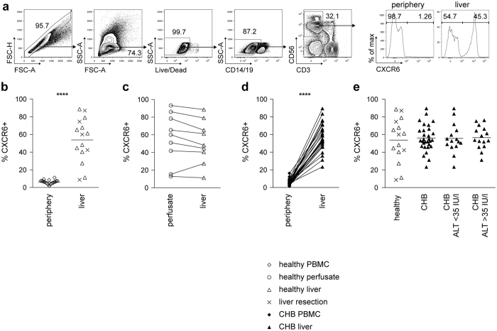
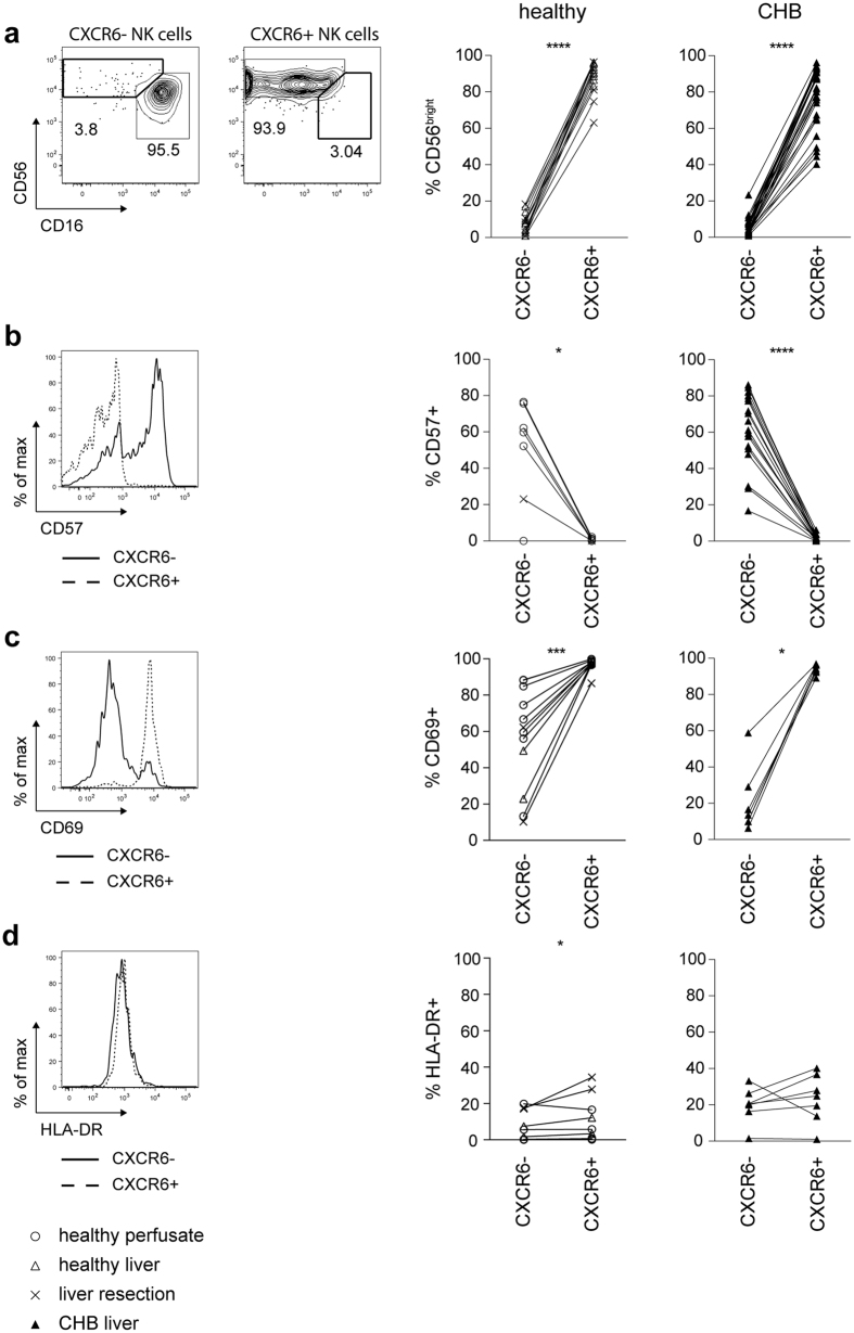
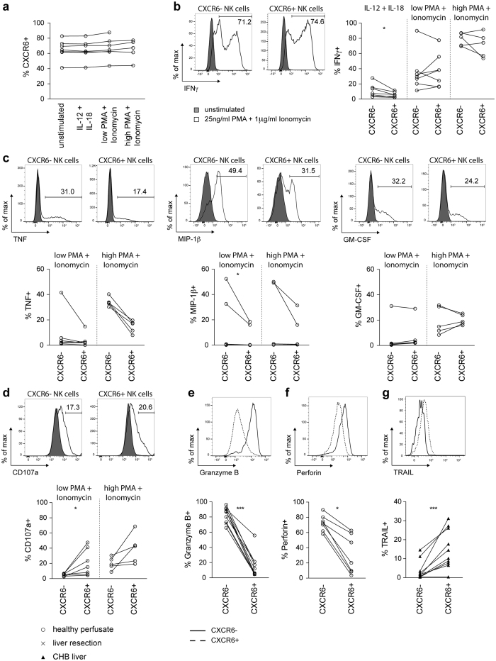
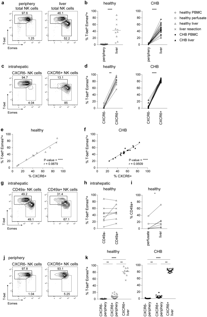
Natural killer cells (NK) are highly enriched in the human liver, where they can regulate immunity and immunopathology. We probed them for a liver-resident subset, distinct from conventional bone-marrow-derived NK. CXCR6+ NK were strikingly enriched in healthy and diseased liver compared to blood (p < 0.0001). Human hepatic CXCR6+ NK had an immature phenotype (predominantly CD56(bright)CD16-CD57-), and expressed the tissue-residency marker CD69. CXCR6+ NK produced fewer cytotoxic mediators and pro-inflammatory cytokines than the non-liver-specific CXCR6- fraction. Instead CXCR6+ NK could upregulate TRAIL, a key death ligand in hepatitis pathogenesis. CXCR6 demarcated liver NK into two transcriptionally distinct populations: T-bet(hi)Eomes(lo)(CXCR6-) and T-bet(lo)Eomes(hi)(CXCR6+); the latter was virtually absent in the periphery. The small circulating CXCR6+ subset was predominantly T-bet(hi)Eomes(lo), suggesting its lineage was closer to CXCR6- peripheral than CXCR6+ liver NK. These data reveal a large subset of human liver-resident T-bet(lo)Eomes(hi) NK, distinguished by their surface expression of CXCR6, adapted for hepatic tolerance and inducible anti-viral immunity.


Figures

Figure 1
Figure 1. CXCR6+ NK cells are enriched in the liver in health and disease.
(a) Gating strategy for identification of NK cells (singlets, live cells, CD14−, CD19−, CD3−, CD56+ cells) using multicolor flow cytometry and representative histograms of CXCR6 staining of NK cells from periphery and liver. (b) Summary frequencies of CXCR6+ NK cells from PBMC (n = 20), and liver (n = 15), comprising biopsies obtained from healthy donor livers (n = 10) or healthy margins of resections for colorectal metastases (n = 5). (c) Direct comparison of CXCR6 frequencies of NK cells isolated from perfusion liquid and liver tissue from the same healthy controls (n = 9). (d) Summary of CXCR6 frequencies of NK cells from paired PBMC and liver biopsies from patients with CHB (n = 30). (e) Summary of CXCR6 expression on NK cells from healthy liver (n = 15), compared to liver tissue from patients with CHB (n = 30). CHB patients were additionally divided by ALT (low ALT < 35 IU/l, n = 15; high ALT > 35 IU/l, n = 15). Diagrams show individual data for healthy controls and CHB. P-values indicated in each diagram. Mann-Whitney U-test (b), Kruskal-Wallis with Dunn’s multiple comparison test (e) and paired Wilcoxon test (c,d) were applied. ****p-value < 0.0001.
Figure 2
Figure 2. Intrahepatic CXCR6+ NK cells have an immature phenotype and express high levels of CD69.
(a) Representative contour plots for identification of CD56bright and CD56dim NK cells based on CD16/CD56 expression within intrahepatic CXCR6− and CXCR6+NK cells. Summary of frequency of CD56bright NK cells in intrahepatic CXCR6− and CXCR6+ NK cells of liver tissue of healthy controls (n = 15) and patients with CHB (n = 30). (b) Representative histogram overlay of CD57 expression on intrahepatic CXCR6− and CXCR6+ NK cells. Summary of frequency of CD57 expressing intrahepatic CXCR6− and CXCR6+NK cells of healthy controls (perfusion liquid n = 6; liver tissue n = 1) and patients with CHB (n = 18). (c) Representative histogram overlaying CD69 expression on intrahepatic CXCR6− and CXCR6+ NK cells. Summary of frequency of CD69 expressing intrahepatic CXCR6− and CXCR6+ NK cells of healthy controls (fresh perfusion liquid n = 2; frozen perfusion liquid n = 6; liver tissue n = 4) and of patients with CHB (n = 7). (d) Representative histogram overlaying HLA-DR expression on intrahepatic CXCR6− and CXCR6+ NK cells. Summary of frequency of HLA-DR expressing intrahepatic CXCR6− and CXCR6+ NK cells of healthy controls (fresh perfusion liquid n = 2; frozen perfusion liquid n = 6; liver tissue n = 4) and patients with CHB (n = 7). Paired Wilcoxon test was applied for all figures. *p-value < 0.05, ***p-value < 0.001, ****p-value < 0.0001.
Figure 3
Figure 3. Functional capacity of CXCR6− and CXCR6+ intrahepatic NK cells.
(a) Summary of percentage of NK cells isolated from perfusates expressing CXCR6 with or without stimulation with IL-12/IL-18 (5 ng/ml, 50 ng/ml) or PMA and Ionomycin at low (3 ng/ml, 100 ng/ml) or high (25 ng/ml, 1 μg/ml) dose (n = 7; 7; 7; 5). (b) Representative histograms overlaying IFNγ production of unstimulated, and high dose PMA/Ionomycin stimulated, for intrahepatic CXCR6− and CXCR6+ NK cells. Summary of IFNγ production by CXCR6− and CXCR6+ NK cells from healthy liver perfusates with stimuli as indicated (n = 7; 7; 5), after subtraction of the unstimulated control. (c) Representative histograms overlaying TNF, MIP-1β and GM-CSF production of unstimulated, and high dose PMA/Ionomycin stimulated, intrahepatic CXCR6− and CXCR6+ NK cells. Summary of TNF, MIP-1β and GM-CSF production by CXCR6− and CXCR6+ NK cells from healthy liver perfusates with stimuli as indicated (n = 7; 5), after subtraction of the unstimulated control. (d) Representative histograms overlaying CD107a expression of unstimulated, and high dose PMA/Ionomycin stimulated, intrahepatic CXCR6− and CXCR6+NK cells. Summary of CD107a expression by CXCR6− and CXCR6+NK cells from healthy liver perfusates with stimuli as indicated (n = 7; 5), after subtraction of the unstimulated control. (e) Representative histogram overlaying ex vivo granzyme B expression in intrahepatic CXCR6− and CXCR6+ NK cells. Summary of frequency of granzyme B expressing intrahepatic CXCR6− and CXCR6+ NK cells from healthy controls (perfusion liquid n = 9; liver tissue n = 1). (f) Representative histogram overlaying ex vivo perforin expression in intrahepatic CXCR6− and CXCR6+ NK cells. Summary of frequency of perforin expressing intrahepatic CXCR6− and CXCR6+ NK cells from healthy controls (perfusion liquid n = 7). (g) Representative histogram overlaying TRAIL expression on intrahepatic CXCR6− and CXCR6+ NK cells from a patient with CHB. Summary of frequency of TRAIL expressing intrahepatic CXCR6− and CXCR6+ NK cells from patients with CHB (n = 10). Kruskal-Wallis with Dunn’s multiple comparison test (A) and Paired Wilcoxon test (B–G) was applied. *p-value < 0.05, ***p-value < 0.001.
Figure 4
Figure 4. The liver contains a distinct T-betloEomeshi NK cell subset identified by CXCR6, but not CD49a expression.
(a) Representative contour plots for identification of T-bethiEomeslo and T-betloEomeshi subsets of total NK cells from periphery and liver tissue. (b) Summary of frequency of T-betloEomeshi cells of total NK cells in PBMC (n = 20) and liver tissue of healthy controls (n = 10), and paired PBMC and liver biopsies from patients with CHB (n = 19). (c) Representative contour plots for identification of T-betloEomeshi and T-bethiEomeslo subsets in intrahepatic CXCR6− and CXCR6+ NK cells. (d) Summary of frequency of T-betloEomeshi cells among intrahepatic CXCR6− and CXCR6+ NK cells of liver tissue of healthy controls (n = 10) and patients with CHB (n = 19). (e,f) Correlation of frequency of T-betloEomeshi and CXCR6 expression of NK cells from liver tissue of healthy controls ((e) n = 10) and liver biopsies from patients with CHB ((f) n = 19). (g) Representative contour plots for identification of T-betloEomeshi and T-bethiEomeslo subsets among intrahepatic CD49a− and CD49a+ NK cells. (h) Summary of frequency of T-betlo Eomeshi cells among intrahepatic CD49a− and CD49a+ NK cells of healthy liver tissue (n = 8). (i) Summary of frequency of CD49a expression on intrahepatic NK cells from paired perfusion liquid and tissue from pre-implant donor livers (n = 7). (j) Representative contour plots for identification of T-betloEomeshi and T-bethiEomeslo subsets among peripheral CXCR6− and CXCR6+ NK cells. (k) Summary of frequency of T-betloEomeshi cells among peripheral CXCR6− and CXCR6+ NK cells (n = 20) and intrahepatic CXCR6+NK cells of liver tissue of healthy controls (n = 10); and of peripheral CXCR6− and CXCR6+ NK cells and paired intrahepatic CXCR6+ NK cells of patients with CHB (n = 19). P-values and r indicated in each diagram. Mann-Whitney U-test ((b) left), Kruskal-Wallis with Dunn’s multiple comparison test ((k) left), Friedmans with Dunn’s multiple comparision test ((k) right), paired Wilcoxon test ((b) right, (d,h,i)) and Spearman’s correlation (e,f) were applied. *p-value < 0.05, **p-value < 0.01, ****p-value < 0.0001.

Similar articles

Cited by

References

    1. Doherty D. G. et al.. The human liver contains multiple populations of NK cells, T cells, and CD3+CD56+ natural T cells with distinct cytotoxic activities and Th1, Th2, and Th0 cytokine secretion patterns. J Immunol 163, 2314–2321 (1999). - PubMed
    1. Maini M. K. & Peppa D. NK cells: a double-edged sword in chronic hepatitis B virus infection. Front Immunol 4, 57, doi: 10.3389/fimmu.2013.00057 (2013). - DOI - PMC - PubMed
    1. Peppa D. et al.. Blockade of immunosuppressive cytokines restores NK cell antiviral function in chronic hepatitis B virus infection. PLos Pathog 6, e1001227, doi: 10.1371/journal.ppat.1001227 (2010). - DOI - PMC - PubMed
    1. Dunn C. et al.. Cytokines induced during chronic hepatitis B virus infection promote a pathway for NK cell-mediated liver damage. J Exp Med 204, 667–680, doi: 10.1084/jem.20061287 (2007). - DOI - PMC - PubMed
    1. Micco L. et al.. Differential boosting of innate and adaptive antiviral responses during pegylated-interferon-alpha therapy of chronic hepatitis B. J Hepatol 58, 225–233, doi: 10.1016/j.jhep.2012.09.029 (2013). - DOI - PubMed

Publication types

MeSH terms