. 2016 May 23:6:26669.
doi: 10.1038/srep26669.

Predominance of sperm motion in corners

Affiliations

Predominance of sperm motion in corners

Reza Nosrati et al. Sci Rep. .

Abstract

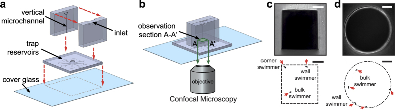
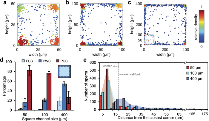
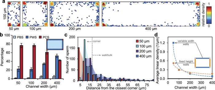
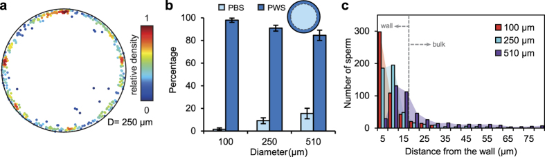
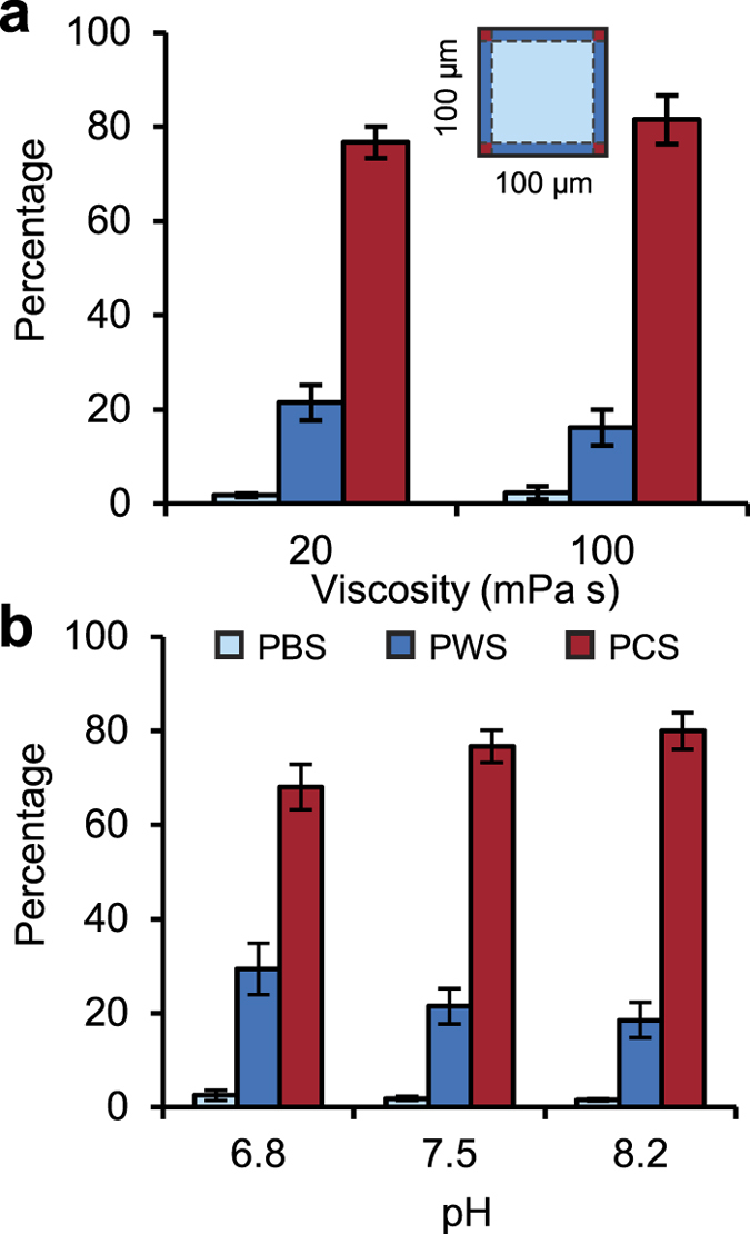
Sperm migration through the female tract is crucial to fertilization, but the role of the complex and confined structure of the fallopian tube in sperm guidance remains unknown. Here, by confocal imaging microchannels head-on, we distinguish corner- vs. wall- vs. bulk-swimming bull sperm in confined geometries. Corner-swimming dominates with local areal concentrations as high as 200-fold that of the bulk. The relative degree of corner-swimming is strongest in small channels, decreases with increasing channel size, and plateaus for channels above 200 μm. Corner-swimming remains predominant across the physiologically-relevant range of viscosity and pH. Together, boundary-following sperm account for over 95% of the sperm distribution in small rectangular channels, which is similar to the percentage of wall swimmers in circular channels of similar size. We also demonstrate that wall-swimming sperm travel closer to walls in smaller channels (~100 μm), where the opposite wall is within the hydrodynamic interaction length-scale. The corner accumulation effect is more than the superposition of the influence of two walls, and over 5-fold stronger than that of a single wall. These findings suggest that folds and corners are dominant in sperm migration in the narrow (sub-mm) lumen of the fallopian tube and microchannel-based sperm selection devices.


Figures

Figure 1
Figure 1. Microfluidic device for quantifying cross-sectional distribution of sperm in microchannels.
(a) Schematic view of the device: a microchannel is vertically aligned with an observation chamber in the horizontal layer. (b) A unique head-on microchannel confocal microscopy approach was used for imaging. The shallow focal plane was focused at the entry of the vertical microchannel to the observation chamber and then moved 20 μm inside the channel. A representative bright-field image of the microchannel cross-section (top) and fluorescence image of sperm in the microchannel cross-section (bottom) for (c) rectangular and (d) circular microchannels. White dash lines in fluorescence images indicate the microchannel wall, acquired using bright-field images, and red arrows point to sperm. Scale bars represent 30 μm and 45 μm for rectangular and circular microchannels, respectively.
Figure 2
Figure 2. Corner-swimming preference of sperm in square microchannels.
Cross-sectional distribution of 1176, 1457, and 1188 bull sperm in square microchannels with side-lengths of (a) 50 μm, (b) 100 μm, and (c) 400 μm, respectively. For comparison, the 50 μm and 100 μm channels are shown to scale inset in the bottom left corner of the 400 μm channel plot. The color bar represents the relative density of sperm in each graph. (d) Percentage of bulk swimmer (PBS), percentage of wall swimmer (PWS), and percentage of corner swimmer (PCS) sperm as a function of microchannel size. Wall-swimming sperm values do not include corner swimmers. Each point represents experiments with at least three samples with a minimum of 874 sperm imaged in each experiment. Values are reported as mean ± s.d. (e) Histograms of sperm distance from the closest corner for each case. A total number of 800 sperm is included in each case.
Figure 3
Figure 3. Corner-swimming preference of sperm in rectangular microchannels with varying width, all 100 μm in height.
(a) Cross-sectional distribution of 929, 1068, 1023, and 956 bull sperm in rectangular microchannels of 100 μm in height and 50 μm, 100 μm, 200 μm, and 400 μm in width. The color bar represents the relative density of sperm in each graph. (b) PBS, PWS and PCS sperm as a function of microchannel width. Wall-swimming sperm do not include corner-swimming sperm. Each bar represents at least three experiments with a minimum of 589 sperm imaged in each. Values are reported as mean ± s.d. (c) Histograms of sperm distance from the closest corner for all rectangular microchannels. A total number of 500 sperm is included in each case. (d) Average linear density - percentage of wall swimmers per unit length of wall – plotted for both the fixed height walls and the variable width walls, for all four cases.
Figure 4
Figure 4. Swimming preference of sperm in circular microchannels.
(a) A representative cross-sectional distribution of 596 bull sperm in circular microchannels of 250 μm in diameter. Color bar represents the relative density of sperm in each graph. (b) PBS and PWS sperm as a function of microchannel diameter. Each points represents experiments with at least three samples with a minimum of 494 sperm imaged in each of the experiments. Values are reported as mean ± s.d. (c) Histograms of sperm distance from the wall for all circular microchannels. A total number of 494 sperm is included in each case.
Figure 5
Figure 5. Effects of the swimming medium on corner-swimming preference of sperm in 100 × 100 μm microchannels.
PBS, PWS, and PCS sperm as a function of (a) viscosity and (b) pH of swimming media. Wall-swimming sperm do not include corner-swimming sperm. Each bar represents at least three experiments with a minimum of 874 sperm imaged in each. Values are reported as mean ± s.d.

References

    1. Miki K. & Clapham D. E. Rheotaxis guides Mammalian sperm. Curr. Biol. 23, 443–452 (2013). - PMC - PubMed
    1. Smith D. J., Gaffney E. A., Gadelha H., Kapur N. & Kirkman-Brown J. C. Bend propagation in the flagella of migrating human sperm, and Its modulation by viscosity. Cell Motil. Cytoskeleton 66, 220–236 (2009). - PubMed
    1. Smiljakovic T. et al.. The role of pH values in porcine reproductive tracts of male and female individuals. Biotechnol. Anim. Husb. 24, 101–108 (2008).
    1. Eisenbach M. & Giojalas L. C. Sperm guidance in mammals - an unpaved road to the egg. Nat. Rev. Mol. Cell Biol. 7, 276–285 (2006). - PubMed
    1. Suarez S. S. & Pacey A. A. Sperm transport in the female reproductive tract. Human Reproduction Update 12, 23–37 (2006). - PubMed

Publication types

Grants and funding