Review
. 2016 Jul;12(7):383-401.
doi: 10.1038/nrneph.2016.70. Epub 2016 May 23.

Complement in disease: a defence system turning offensive

Affiliations
Review

Complement in disease: a defence system turning offensive

Daniel Ricklin et al. Nat Rev Nephrol. 2016 Jul.

Abstract

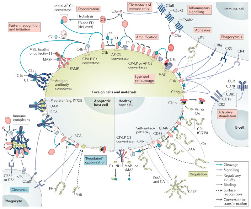
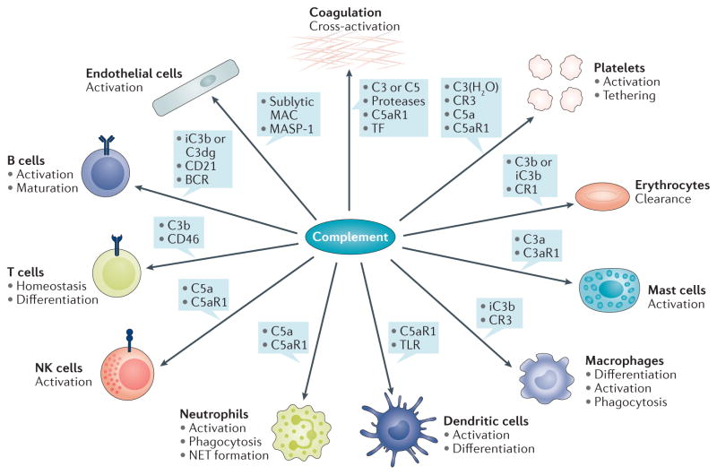
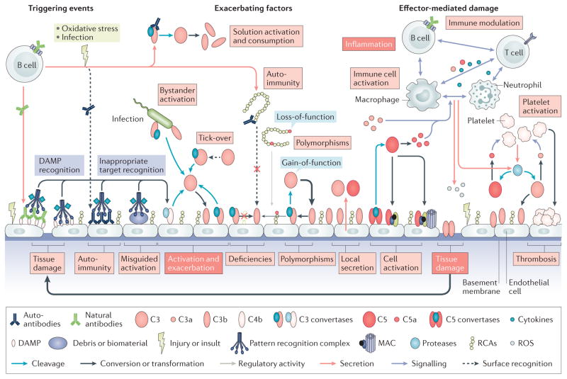
Although the complement system is primarily perceived as a host defence system, a more versatile, yet potentially more harmful side of this innate immune pathway as an inflammatory mediator also exists. The activities that define the ability of the complement system to control microbial threats and eliminate cellular debris - such as sensing molecular danger patterns, generating immediate effectors, and extensively coordinating with other defence pathways - can quickly turn complement from a defence system to an aggressor that drives immune and inflammatory diseases. These host-offensive actions become more pronounced with age and are exacerbated by a variety of genetic factors and autoimmune responses. Complement can also be activated inappropriately, for example in response to biomaterials or transplants. A wealth of research over the past two decades has led to an increasingly finely tuned understanding of complement activation, identified tipping points between physiological and pathological behaviour, and revealed avenues for therapeutic intervention. This Review summarizes our current view of the key activating, regulatory, and effector mechanisms of the complement system, highlighting important crosstalk connections, and, with an emphasis on kidney disease and transplantation, discusses the involvement of complement in clinical conditions and promising therapeutic approaches.


Conflict of interest statement

Competing interests statement

J.D.L and D.R. are inventors of patents or patent applications that describe the use of complement inhibitors for therapeutic purposes. J.D.L. is the founder of Amyndas Pharmaceuticals, which is developing complement inhibitors. E.S.R. declares no competing interests.

Figures

Figure 1
Figure 1. Mechanisms of differential complement activation and regulation under physiological conditions
When complement encounters a foreign cell, the recognition molecules C1q, mannose-binding lectin (MBL), ficolins, and collectin-11 sense antibody clusters or pathogen-associated molecular patterns (PAMPs) and activate associated serine proteases (C1r, C1s or MBL-associated serine proteases (MASPs)) that cleave C4 and C2 to form the C3 convertase (C4b2b) of the classical pathway (CP) and lectin pathway (LP) on the triggering cell. In addition, tick-over or surface interactions of C3 with certain cell surfaces leads to C3 hydrolysis and generation of initial alternative pathway (AP) C3 convertases. All C3 convertases cleave C3 into the anaphylatoxin C3a and C3b, the latter of which is deposited on activating surfaces (opsonization). Factor B (FB) binds to C3b and gets activated by Factor D (FD) to form the final AP convertases (C3bBb), which activate more C3 to C3b and fuel an amplification loop. Increasing densities of C3b deposition favour the generation of C5 convertases, which activate C5 to release the anaphylatoxin C5a and C5b, which, after tiered interactions with C6–C9 and membrane insertion, in turn forms membrane attack complexes (MACs), which leads to lysis, damage, or activation of target cells. The released anaphylatoxins act as immune mediators and, particularly in the case of C5a, attract and prime immune cells. Interaction of the C3b opsonin and its degradation products iC3b and C3dg with complement receptors (CRs) mediates cell adhesion (via CR1 (also known as CD35)) and/or phagocytic uptake (via CR3, CR4, and CRIg). Concurrently, iC3b and C3dg modulate adaptive immune responses by binding to CR2 on the B cell co receptor, thereby lowering the threshold of B cell stimulation. Properdin (FP) stabilizes the C3 and C5 convertases and increases complement-mediated opsonization and effector generation. On healthy cells, complement activation is tightly controlled. C1 esterase inhibitor (C1-INH), MAP1 and sMAP regulate the activity of recognition complexes, whereas soluble C4b-binding protein (C4BP) and factor H (FH) — which recognize self-surface patterns such as heparin and sialic acid, and bind to host cells — or membrane-bound regulators of complement activation (RCA) proteins (CD35, CD46 and CD55) control convertase activity by accelerating convertase decay and/or act as cofactors for the factor I (FI)-mediated degradation of C3b and C4b. Finally, CD59, clusterin (clu) and vitronectin (vtn) prevent formation of MAC. ‘Silent’ removal of damaged and apoptotic cells, debris, and immune complexes is achieved through the sensing of damage-associated molecular patterns (DAMPs), either directly by complement recognition complexes or mediated by modulators such as pentraxins (for example, PTX3). Controlled activation of complement occurs with limited deposition of opsonins that facilitate phagocytic clearance. In addition to C3b/iC3b/C3dg, C1q also contributes to clearance by binding to C1q receptors (gC1qR and cC1qR). CA, cofactor activity; CRIg, CR of the immunoglobulin superfamily; DAA, decay acceleration activity; FcγR, Fcγ receptor.
Figure 2
Figure 2. Examples of complement crosstalk with immune cells and defence pathways
The generation of complement effectors stimulates a broad spectrum of downstream immune, inflammatory, and procoagulative responses. The anaphylatoxin C5a, for example, exerts strong proinflammatory effects by acting as a chemoattractant and stimulator of various immune cells via C5aR1-mediated signalling, thereby influencing priming and activation with release of mediators (for example, cytokines, neutrophil extracellular traps (NETs)), differentiation, and functional activity. C3a has a distinct spectrum from C5a and has, for example, been shown to activate mast cells. Activation of professional phagocytes induces the expression of complement receptors, which enable complement-mediated phagocytosis, whereas crosstalk between C5aR, FcγR, and dectin-1 also affects antibody-mediated uptake. Adherence of opsonins to CR1 on erythrocytes is an important mechanism that directs immune complexes to the liver and spleen. Complement activation also modulates adaptive immune responses by lowering the threshold of B-cell stimulation (via iC3b or C3dg interaction with CD21) or by influencing T-cell activation (for example, by binding of C3b to CD46), differentiation, and homeostasis. Complement effectors such as C5a, sublytic membrane attack complex (MAC), and MASP-1, can directly activate endothelial cells and, for example, increase expression of tissue factor (TF) as an inducer of coagulation. Serine proteases of the complement and coagulation systems might cross-activate under certain circumstances to contribute to thrombo-inflammation. Concomitantly, the release of complement proteins and binding of both complement activators and regulators to platelets might amplify the platelet response and contribute to clearance of platelets and pathogens alike. BCR, B-cell receptor; NK, natural killer; TLR, Toll-like receptor.
Figure 3
Figure 3. Pathological activation of complement
Various events contribute to the development and progression of complement-related diseases. Excessive generation of triggers, such as the massive influx of pathogen-associated molecular patterns during sepsis or release of damage-associated molecular patterns (DAMPs) during trauma or tissue injury, leads to binding of recognition molecules and induces a complement response. These pronounced complement responses can lead to bystander activation of healthy host tissues in proximity to the initial trigger (for example, a pathogen). Moreover, the recognition of inappropriate targets, such as biomaterials (for example, implants or haemodialysis filters) or accumulating debris resulting from ageing or oxidative stress can induce misguided complement activation that contributes to inflammatory complications. Complement deficiencies can contribute to the generation of autoantibodies owing to impaired removal of immune complexes and other mechanisms, and the resulting antibodies can activate complement by binding to neoantigens on damaged cells or to self-antigens on healthy cells, with subsequent sensing by C1 complexes. Autoantibodies can also influence the activity of the complement system by interfering with the surface-recognition capacity of regulators of complement activation (RCA) proteins such as factor H (FH), or by stabilizing convertase complexes, thereby exacerbating complement activation. Gain-of-function mutations in complement components or loss-of-function mutations and deficiencies in complement regulators largely define the systemic activation profile of complement (marked by thick and thin arrows to indicate the level of activity), and can lead to attack of susceptible organs such as the kidney. In addition to circulating systemic complement, local secretion of complement components by tissue cells and infiltrating or tissue-resident immune cells contributes to activation events. The generation of complement effectors leads to the attraction and activation of immune cells, with release of proinflammatory mediators such as cytokines, reactive oxygen species (ROS) or reactive nitrogen species. The resulting cell damage further stimulates complement activation and fuels a vicious cycle of complement activation, inflammation, and tissue damage. Finally, strong crosstalk between complement and the coagulation system contributes to thrombotic events.

References

    1. Ricklin D, Hajishengallis G, Yang K, Lambris JD. Complement: a key system for immune surveillance and homeostasis. Nat Immunol. 2010;11:785–797. - PMC - PubMed
    1. Ricklin D, Lambris JD. Complement in immune and inflammatory disorders: pathophysiological mechanisms. J Immunol. 2013;190:3831–3838. - PMC - PubMed
    1. Reis ES, et al. Applying complement therapeutics to rare diseases. Clin Immunol. 2015;161:225–240. - PMC - PubMed
    1. Merle NS, Church SE, Fremeaux-Bacchi V, Roumenina LT. Complement system part I — molecular mechanisms of activation and regulation. Front Immunol. 2015;6:262. - PMC - PubMed
    1. Merle NS, Noe R, Halbwachs-Mecarelli L, Fremeaux-Bacchi V, Roumenina LT. Complement system part II: role in immunity. Front Immunol. 2015;6:257. - PMC - PubMed

Publication types

Substances