

TGF-β1-induced PAI-1 contributes to a profibrotic network in patients with eosinophilic esophagitis
- PMID: 27212082
- PMCID: PMC5014565
- DOI: 10.1016/j.jaci.2016.02.028
TGF-β1-induced PAI-1 contributes to a profibrotic network in patients with eosinophilic esophagitis
Abstract
Background: Eosinophilic esophagitis (EoE) is an allergic disease of increasing worldwide incidence. Complications are due to tissue remodeling and involve TGF-β1-mediated fibrosis. Plasminogen activator inhibitor 1 (PAI-1/serpinE1) can be induced by TGF-β1, but its role in EoE is not known.
Objective: We sought to understand the expression and role of PAI-1 in patients with EoE.
Methods: We used esophageal biopsy specimens and plasma samples from control subjects and patients with EoE, primary human esophageal epithelial cells, and fibroblasts from patients with EoE in immunohistochemistry, quantitative PCR, and immunoassay experiments to understand the induction of PAI-1 by TGF-β1, the relationship between PAI-1 and esophageal fibrosis, and the role of PAI-1 in fibrotic gene expression.
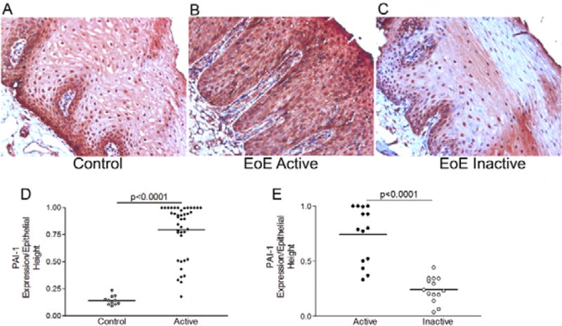
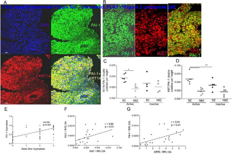
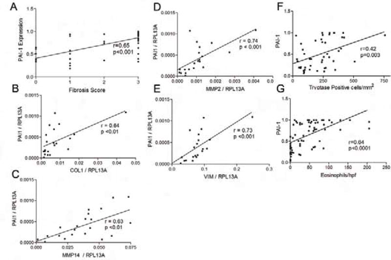
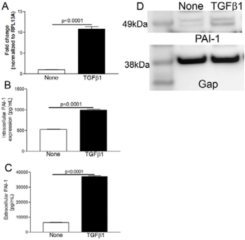
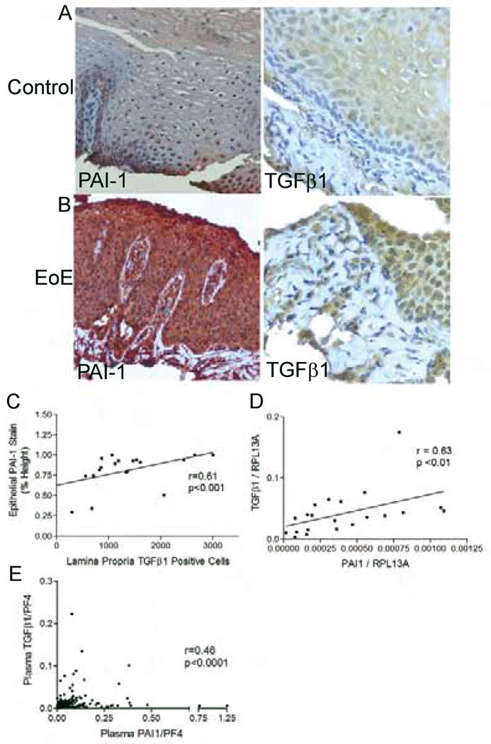
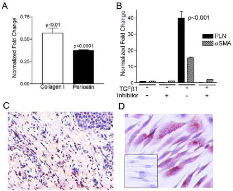
Results: PAI-1 expression was significantly increased in epithelial cells of biopsy specimens from patients with active EoE compared with that seen in biopsy specimens from patients with inactive EoE or control subjects (P < .001). Treatment of primary esophageal epithelial cells with recombinant TGF-β1 increased PAI-1 transcription, intracellular protein expression, and secretion. Esophageal PAI-1 expression correlated with basal zone hyperplasia, fibrosis, and markers of esophageal remodeling, including vimentin, TGF-β1, collagen I, fibronectin, and matrix metalloproteases, and plasma PAI-1 levels correlated with plasma TGF-β1 levels. PAI-1 inhibition significantly decreased baseline and TGF-β1-induced fibrotic gene expression.
Conclusions: PAI-1 expression is significantly increased in the epithelium in patients with EoE and reflects fibrosis, and its inhibition decreases TGF-β1-induced gene expression. Epithelial PAI-1 might serve as a marker of EoE severity and form part of a TGF-β1-induced profibrotic network.
Keywords: Eosinophil; SerpinE1; TGF-β1; esophagitis; fibrosis; remodeling.
Copyright © 2016 American Academy of Allergy, Asthma & Immunology. Published by Elsevier Inc. All rights reserved.
Figures
References
-
- Liacouras CA, Furuta GT, Hirano I, Atkins D, Attwood SE, Bonis PA, et al. Eosinophilic esophagitis: updated consensus recommendations for children and adults. The Journal of allergy and clinical immunology. 2011 Jul;128(1):3–20. e6. quiz 1–2. Epub 2011/04/12. eng. - PubMed
-
- Schoepfer AM, Safroneeva E, Bussmann C, Kuchen T, Portmann S, Simon HU, et al. Delay in diagnosis of eosinophilic esophagitis increases risk for stricture formation in a time-dependent manner. Gastroenterology. 2013 Dec;145(6):1230–6. e1–2. - PubMed
-
- Aceves SS, Newbury RO, Dohil R, Bastian JF, Broide DH. Esophageal remodeling in pediatric eosinophilic esophagitis. The Journal of allergy and clinical immunology. 2007 Jan;119(1):206–12. - PubMed
Publication types
MeSH terms
Substances
Grants and funding
LinkOut - more resources
Full Text Sources
Other Literature Sources
Medical
Miscellaneous
