. 2016 Jun 16;165(7):1658-1671.
doi: 10.1016/j.cell.2016.04.049. Epub 2016 May 19.

Corpse Engulfment Generates a Molecular Memory that Primes the Macrophage Inflammatory Response

Affiliations

Corpse Engulfment Generates a Molecular Memory that Primes the Macrophage Inflammatory Response

Helen Weavers et al. Cell. .

Abstract

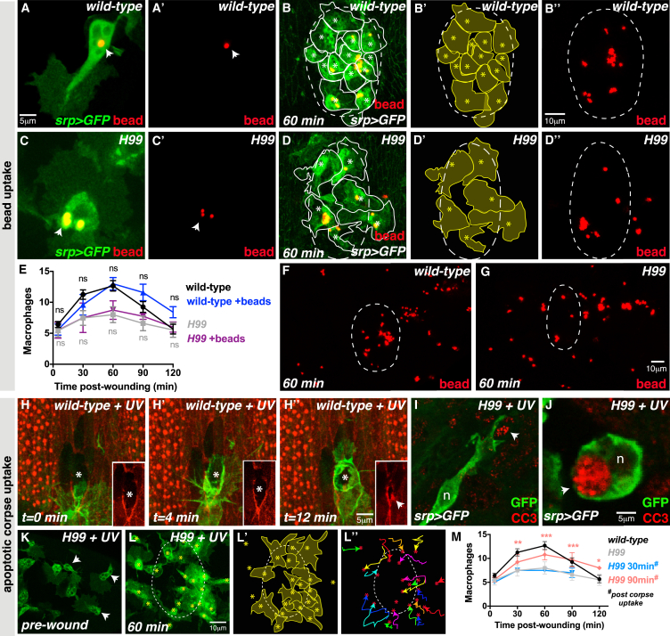
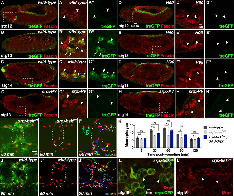
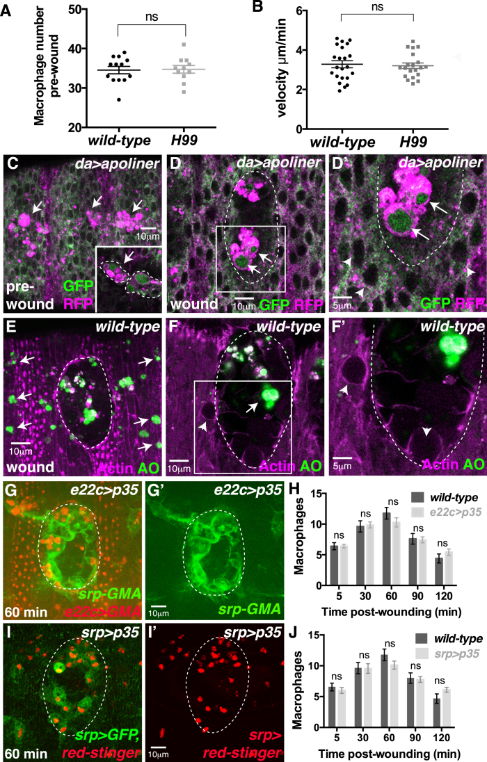
Macrophages are multifunctional cells that perform diverse roles in health and disease. Emerging evidence has suggested that these innate immune cells might also be capable of developing immunological memory, a trait previously associated with the adaptive system alone. While recent studies have focused on the dramatic macrophage reprogramming that follows infection and protects against secondary microbial attack, can macrophages also develop memory in response to other cues? Here, we show that apoptotic corpse engulfment by Drosophila macrophages is an essential primer for their inflammatory response to tissue damage and infection in vivo. Priming is triggered via calcium-induced JNK signaling, which leads to upregulation of the damage receptor Draper, thus providing a molecular memory that allows the cell to rapidly respond to subsequent injury or infection. This remarkable plasticity and capacity for memory places macrophages as key therapeutic targets for treatment of inflammatory disorders.


Figures

None
Graphical abstract
Figure 1
Figure 1
Diverse Macrophage Strategies Clear Dying Apoptotic Cells In Vivo (A–I) Drosophila macrophages migrate along the ventral nerve cord (VNC) (arrows, A) and engulf apoptotic cells (B). Naive macrophages (lacking corpses, C) engulf corpses (asterisks) at close range (arrow, D) or using long pseudopods (arrow, E). Trailing macrophages reach outlying corpses (arrow, F) by migration off the midline (dashed line). Uptake strategies are quantified in (G). Corpses accumulate as cytoplasmic vacuoles (arrowheads, inset H; quantified in I). Macrophages reach the three-row arrangement by stage 15 (arrows, H). Macrophages labeled using srp-Gal4 driving UAS-red-stinger (nuclei, red) and UAS-GFP (cytoplasm, green). (J–K′) Apoptotic corpses detected in macrophages (green, srp >GFP; nuclear DAPI, blue) using cleaved caspase-3 (CC3, red; arrows, J and J′) or the Apoliner caspase sensor (driven ubiquitously by daughterless-Gal4; uncleaved Apoliner, red; cleaved nuclear Apoliner, green; arrows, K and K′). See also Movie S1.
Figure 2
Figure 2
Apoptotic Corpses Prime Macrophages for Detection of Tissue Damage (A–H) H99 macrophages (srp-Gal4 driving red-stinger and GFP) do not encounter corpses (lack of cytoplasmic vacuoles, A) but migrate normally (B) and reach the characteristic three-row arrangement (arrows, C). H99 macrophages are not robustly recruited to wounds (D–D′′, quantified in E) unlike wild-type macrophages (F–F′′; arrows in G indicate corpses of wild-type macrophage). Data are represented as mean ± SEM; ns, not significant; ∗∗p < 0.01 and ∗∗∗p < 0.001 via one-way ANOVA followed by Sidak’s multiple comparisons test (E). H99 macrophages within the wound (dashed line, H) phagocytose necrotic debris (asterisks, H); macrophages outside the wound (arrows, H) extend pseudopods to engulf wound debris (arrowheads, H). (I and J) Wound H2O2 production (Amplex Red, red) is indistinguishable from wild-type (I) in H99 mutants (J). Macrophages labeled using srp >GFP (green). (K–L′) Apoptotic cells (anti-CC3, blue) are not detected in the wild-type wounded epithelium (K; actin, red). Macrophages outside the wound (dashed outlines, K) and within the wound (dashed outlines, L and L′) contain corpses engulfed earlier during dispersal (arrows, insets L and L′). See also Figure S1 and Movies S2 and S3.
Figure 3
Figure 3
Naive Macrophages Are Experimentally Primed by Corpse Uptake (A–G) Wild-type (A and A′) and H99 (C and C′) macrophages (srp >GFP, green) engulf beads (red; arrows). Wild-type macrophages (yellow outlines, B and B′) with beads (red, high magnification in B′′ and low magnification in F) are robustly recruited to wounds (B–B′′ and E) but H99 macrophages (yellow outlines, D and D′) with beads (D′′ and low magnification in G) are not (D–D′′ and E). (H–M) UV-induced apoptosis triggered in a single cell (asterisk) that assembles a cortical actin cable and delaminates (arrow) from epithelium (actin, red; inset H–H′′). Macrophage (green, srp >GFP) engulfs apoptotic cell (H′ and H′′). UV-triggered corpses detected in H99 mutants by anti-CC3 (red; arrows) during (I) and after (J) uptake. Corpse uptake by H99 macrophages (green, srp > GFP; pre-wound, K) rescues the wound recruitment defect (macrophages marked by asterisks; outlined in L′) for wounds made 90 min, but not 30 min, post-corpse uptake (L–L′′ and M). For (E) and (M), data are represented as mean ± SEM; ns, not significant; p < 0.05, ∗∗p < 0.01, and ∗∗∗p < 0.001 via one-way ANOVA followed by Sidak’s multiple comparisons test. Significance shown for H99 90-min treatment compared to H99 untreated in (M).
Figure 4
Figure 4
Corpse-Induced Calcium Bursts Prime Macrophages for Wound Recruitment (A–F′′) Wild-type macrophages exhibit rapid calcium flashes (arrowheads; green, srp-Gal4>UAS-GCaMP3) upon corpse uptake (A, inset A′ and B). A single calcium flash occurs upon each engulfment (first engulfment in C, insets D–D′′; second engulfment by same cell 3 min later in E, insets F–F′′). Macrophage nuclei (red) labeled using srp-Gal4 >UAS-red-stinger. (G and H) Inhibition of calcium bursts (srp-Gal4>UAS-parvalbumin) impairs macrophage migration to wounds (G and G′; quantified in H). Macrophages labeled using srp-Gal4 driven red-stinger (nuclei, red) and GFP (cytoplasm, green). Data are represented as mean ± SEM; ns, not significant; p < 0.05 and ∗∗∗p < 0.001 via one-way ANOVA followed by Sidak’s multiple comparisons test (H). See also Figure S2 and Movies S4 and S5.
Figure 5
Figure 5
Corpse-Induced Draper Expression Primes Macrophages (A–E′) Draper transcript (A and B) and protein (C and E) levels increase upon corpse uptake. Naive stage 11 macrophages have low levels of Draper transcripts (arrowheads, A) and protein (arrowheads, C and C′) that increase after corpse uptake (D and E); Draper protein relocalizes from corpse-associated punctae (arrowheads, D and D′′) to the cell cortex (arrowheads, E′). (F–L′′) Stage 15 H99 macrophages have low Draper transcript (arrowheads, F) and protein (arrowheads, G and G′) levels but Draper expression is increased 90 min after UV-induced corpse uptake (arrowheads, H and H′). Inhibition of macrophage calcium signaling also disrupts Draper expression (arrowheads, I). Ectopic Draper expression in H99 macrophages (driven by srp-Gal4) restores macrophage wound recruitment (J–J′′ and K) to wild-type levels (L–L′′). For (K), data are represented as mean ± SEM; ns, not significant; ∗∗p < 0.01 and ∗∗∗p < 0.001 via one-way ANOVA followed by Sidak’s multiple comparisons test. See also Figure S3.
Figure 6
Figure 6
Corpse-Induced JNK Signaling Primes Macrophages (A–H′′) JNK activity (green, treGFP reporter) is absent from naive macrophages (red, anti-Fascin) at stage 12 (arrowheads; A–A′′). JNK activity increases as macrophages engulf corpses; JNK activity is initially mosaic (B–B′′) and detected in some macrophages (arrows) but not others (arrowheads) but later spreads to all macrophages (arrows, C–C′′). JNK activity is not detected in naive H99 macrophages (arrowheads, D–F′′) or following inhibition of calcium signaling (arrowheads, G–H′′). (I–L′) Inhibition of JNK signaling (srp > bskDN) impairs the wound inflammatory response (compare I–I′′ with wild-type in J–J′′; quantified in K) and disrupts Draper expression (red; arrowheads, L and L′), but wound recruitment is rescued by ectopic Draper expression (K). Macrophages were labeled using cytoplasmic GFP (I, J, and L) and nuclear Red-Stinger (I and J). For (K), data are represented as mean ± SEM; ns, not significant; ∗∗p < 0.01 and ∗∗∗p < 0.001 via one-way ANOVA followed by Sidak’s multiple comparisons test. See also Figure S4.
Figure 7
Figure 7
Corpse-Induced Calcium and JNK Signaling also Prime Macrophages for Infection (A–K) Wild-type macrophages (green, srp >GFP) engulf RFP-E. coli (arrowheads, red; A, insets B–B′′) or pHrodo-E. coli (red; C–C′′). Naive stage 10 macrophages do not engulf RFP-E. coli (arrowheads, D), but RFP-E. coli is taken up by mature stage 11 macrophages (arrowheads, E). H99 macrophages fail to phagocytose E. coli (F and G) or pHrodo-E. coli (H); RFP-E. coli cluster at the macrophage surface (arrowhead, F) but are not stably bound or engulfed (blue E. coli track, G). Bead engulfment (I; arrow, yellow) does not rescue the H99 bacterial uptake defect (I, blue E. coli track; arrowhead, RFP-E. coli), but phagocytosis of UV-induced apoptotic corpses (asterisks, J) does rescue uptake (arrowhead, J). H99 macrophages that lack corpses in the UV-treated embryo fail to engulf RFP-E. coli (arrowhead, K; blue, E. coli track). (L–P) Inhibition of calcium signaling (L; srp >parvalbumin) or JNK activity (M; srp > bskDN) inhibits macrophage (srp > GFP) uptake of RFP-E. coli (arrowheads; blue E. coli tracks). Ectopic Draper expression in H99 macrophages (driven by srp-Gal4) rescues uptake of RFP-E. coli (arrowheads, N) and pHrodo-E. coli (arrowheads, O and O′). E. coli uptake is quantified in (P) (data are represented as mean ± SEM; ∗∗∗p < 0.001 via one-way ANOVA followed by Sidak’s multiple comparisons test). See also Movie S6.
Figure S1
Figure S1
Apoptotic Cell Death Is Not Detected at Wild-Type Wounds, and Apoptotic Caspases Are Not Required in Macrophages for Wound Recruitment, Related to Figure 2 (A and B) The pre-wound H99 macrophage number (A) and H99 macrophage developmental migration speed (B) are indistinguishable from wild-type. (C–H) Apoptosis is not detected in the damaged epithelium of wild-type wounds (dashed outline) using the Apoliner caspase sensor (pre-wound in C; wounded, D and inset D′) or Acridine Orange (AO; z-stack projection in E and single section F and inset F′). Apoptosis is not detected in the damaged cells at the wound edge (arrowheads in D′ and F′). Macrophages in unwounded embryos contain apoptotic cells engulfed during prior developmental dispersal (arrows, C and inset; nuclear cleaved Apoliner, green) and these remain following epithelial wounding, observed in macrophages both outside (arrows, E) and inside the wound site (arrows, D and F). Inhibition of apoptotic cell death in the wounded epithelium (red, e22c-Gal4 driven expression of UAS-p35) does not affect macrophage (green, srp-GMA) wound recruitment (G and H). (I and J) Macrophage specific expression (using srp-Gal4) of the pan-caspase inhibitor p35 did not affect macrophage wound recruitment (srp-Gal4 > UAS-GFP, red-stinger; I-I′ and quantified in J). All data are represented as mean ± SEM; ns, not significant via Mann-Whitney test (A and B) or one-way ANOVA followed by Sidak’s multiple comparisons test (H and J).
Figure S2
Figure S2
Intracellular Calcium Flashes Are Not Required for Macrophage Specification or Developmental Migration and Are Not Induced by Bead Uptake, Related to Figure 4 (A–C) Inhibition of macrophage calcium signaling (srp-Gal4 driven expression of UAS-parvalbumin) does not affect pre-wound macrophage numbers (A), developmental migration speeds (B) or apoptotic corpse uptake (C) compared to wild-type. (D) Intracellular calcium flashes (srp-Gal4>UAS-gcamp3) are not detected during macrophage phagocytosis of fluorescent beads (arrows, yellow). Macrophages labeled using srp-Gal4 driven expression of UAS-red-stinger (red nuclei). Data are represented as mean ± SEM; ns, not significant via Mann-Whitney test (A-C).
Figure S3
Figure S3
Draper Is Not Required for Bead Uptake, and Draper-Rescued H99 Mutants Have Normal Macrophage Numbers and Migration Speeds, Related to Figure 5 (A and B) Absence of specific signal using control sense drpr in situ probe on wild-type stage 15 embryos. (C and C′) drprΔ5 mutant macrophages (green, srp>GFP) phagocytose beads (red) normally (arrows). (D and E) Ectopic Draper expression in H99 mutant macrophages does not affect pre-wound macrophage numbers (D) or developmental migration speeds (E). All data are represented as mean ± SEM; ns, not significant via Mann-Whitney test (D and E).
Figure S4
Figure S4
JNK-Inhibited Macrophages Exhibit Normal Pre-wound Numbers, Migration Speeds, and Corpse Uptake, Related to Figure 6 (A–C) Inhibition of JNK signaling in macrophages (srp-Gal4 driven expression of UAS-bskDN) does not affect pre-wound macrophage numbers (A), developmental migration speeds (B) or corpse uptake (C). Macrophage numbers and migration speeds are not affected by ectopic expression of Draper in JNK-inhibited macrophages (blue, A and B). All data are represented as mean ± SEM; ns, not significant via Mann-Whitney test (A-C).

Comment in

References

    1. Adachi-Yamada T., Nakamura M., Irie K., Tomoyasu Y., Sano Y., Mori E., Goto S., Ueno N., Nishida Y., Matsumoto K. p38 mitogen-activated protein kinase can be involved in transforming growth factor beta superfamily signal transduction in Drosophila wing morphogenesis. Mol. Cell. Biol. 1999;19:2322–2329. - PMC - PubMed
    1. Bardet P.-L., Kolahgar G., Mynett A., Miguel-Aliaga I., Briscoe J., Meier P., Vincent J.-P. A fluorescent reporter of caspase activity for live imaging. Proc. Natl. Acad. Sci. USA. 2008;105:13901–13905. - PMC - PubMed
    1. Bistoni F., Vecchiarelli A., Cenci E., Puccetti P., Marconi P., Cassone A. Evidence for macrophage-mediated protection against lethal Candida albicans infection. Infect. Immun. 1986;51:668–674. - PMC - PubMed
    1. Bistoni F., Verducci G., Perito S., Vecchiarelli A., Puccetti P., Marconi P., Cassone A. Immunomodulation by a low-virulence, agerminative variant of Candida albicans. Further evidence for macrophage activation as one of the effector mechanisms of nonspecific anti-infectious protection. J. Med. Vet. Mycol. 1988;26:285–299. - PubMed
    1. Blok B.A., Arts R.J.W., van Crevel R., Benn C.S., Netea M.G. Trained innate immunity as underlying mechanism for the long-term, nonspecific effects of vaccines. J. Leukoc. Biol. 2015;98:347–356. - PubMed

Publication types