Hetero-site-specific X-ray pump-probe spectroscopy for femtosecond intramolecular dynamics
- PMID: 27212390
- PMCID: PMC4879250
- DOI: 10.1038/ncomms11652
Hetero-site-specific X-ray pump-probe spectroscopy for femtosecond intramolecular dynamics
Abstract
New capabilities at X-ray free-electron laser facilities allow the generation of two-colour femtosecond X-ray pulses, opening the possibility of performing ultrafast studies of X-ray-induced phenomena. Particularly, the experimental realization of hetero-site-specific X-ray-pump/X-ray-probe spectroscopy is of special interest, in which an X-ray pump pulse is absorbed at one site within a molecule and an X-ray probe pulse follows the X-ray-induced dynamics at another site within the same molecule. Here we show experimental evidence of a hetero-site pump-probe signal. By using two-colour 10-fs X-ray pulses, we are able to observe the femtosecond time dependence for the formation of F ions during the fragmentation of XeF2 molecules following X-ray absorption at the Xe site.
Figures


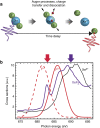
 . In our experiment, after time delays of 4, 29 and 54 fs, a 683-eV probe pulse excites the 1s→2p resonances of F+ or F2+ as the molecule dissociates. (b) Photoabsorption cross-sections for the pump (purple arrow) and probe (red arrow) pulses. The cross-sections are arbitrarily scaled. Neutral XeF2 cross-sections (purple line) are taken from ref. , xenon ions Xeq+ cross-section (black line) are taken from ref. and averaged over q+ using the ion-branching ratios of ref. , and atomic F 1s→2p resonances of F+ or F2+ (red lines) are calculated with Cowan's code. Cross-sections have been broadened to account for the ∼5-eV widths of the ∼10-fs pulses. The two-colour pulses excite the molecule at the Xe site and probe the F ions formed during dissociation.
 . The time delays are 4 fs (black line), 29 fs (green line) and 54 fs (red line). A time dependence is observed for a pump pulse that excites the Xe site at 690 eV and a probe pulse that excites strong 1s→2p resonances near 683 eV of the transient states leading to F+ and F2+ ions. (a,d) Experimental results: the time dependence reveals hetero-site pump-probe events. Pump and probe pulses have ∼10-fs FWHM with ∼5-eV energy widths. (b,c,e,f) Theoretical results based on a classical break-up model for the three time delays, following pathways (1), (2), (3) and (4), respectively. The theoretical results only account for pump-probe events and are consistent with the experimental observations in the KER ranges left from the vertical lines. The error bars represent the standard deviation of the mean.
References
-
- Lépine F., Ivanov M. Y. & Vrakking M. J. J. Attosecond molecular dynamics: fact or fiction? Nature Photon 8, 195–204 (2014).
-
- Zewail A. H. Femtochemistry: atomic-scale dynamics of the chemical bond. J. Phys. Chem. A 104, 5660–5694 (2000). - PubMed
-
- Sundström V. Femtobiology. Annu. Rev. Phys. Chem. 59, 53–77 (2008). - PubMed
-
- Cheng Y.-C. & Fleming G. R. Dynamics of light harvesting in photosynthesis. Ann. Rev. Phys. Chem. 60, 241–262 (2009). - PubMed
Publication types
LinkOut - more resources
Full Text Sources
Other Literature Sources

