

Calcium Sparks in the Heart: Dynamics and Regulation
- PMID: 27212876
- PMCID: PMC4871623
- DOI: 10.2147/RRB.S61495
Calcium Sparks in the Heart: Dynamics and Regulation
Abstract
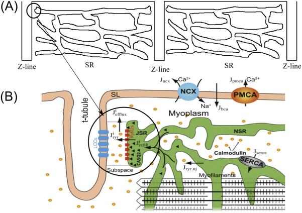
Calcium (Ca2+) plays a central role in the contraction of the heart. It is the bi-directional link between electrical excitation of the heart and contraction. Electrical excitation initiates Ca2+influx across the sarcolemma and T-tubular membrane that triggered calcium release from the sarcoplasmic reticulum. Ca2+sparks are the elementary events of calcium release from the sarcoplasmic reticulum. Therefore, understanding the dynamics of Ca2+sparks is essential for understanding the function of the heart. To this end, numerous experimental and computational studies have focused on this topic, exploring the mechanisms of calcium spark initiation, termination, and regulation and what role these play in normal and patho-physiology. The proper understanding of Ca2+ spark regulation and dynamics serves as the foundation for our insights into a multitude of pathological conditions may develop that can be the result of structural and/or functional changes at the cellular or subcellular level. Computational modeling of Ca2+ spark dynamics has proven to be a useful tool to understand Ca2+ spark dynamics. This review addresses our current understanding of Ca2+ sparks and how synchronized SR Ca2+ release, in which Ca2+ sparks is a major pathway, is linked to the different cardiac diseases, especially arrhythmias.
Keywords: arrhythmia; calcium; heart; sparks.
Figures

Similar articles
-
Regional differences in spontaneous Ca2+ spark activity and regulation in cat atrial myocytes.J Physiol. 2006 May 1;572(Pt 3):799-809. doi: 10.1113/jphysiol.2005.103267. J Physiol. 2006. PMID: 16484302 Free PMC article.
-
Termination of cardiac Ca(2+) sparks: an investigative mathematical model of calcium-induced calcium release.Biophys J. 2002 Jul;83(1):59-78. doi: 10.1016/s0006-3495(02)75149-7. Biophys J. 2002. PMID: 12080100 Free PMC article.
-
Superresolution modeling of calcium release in the heart.Biophys J. 2014 Dec 16;107(12):3018-3029. doi: 10.1016/j.bpj.2014.11.003. Biophys J. 2014. PMID: 25517166 Free PMC article.
-
Excitation-contraction coupling in heart: new insights from Ca2+ sparks.Cell Calcium. 1996 Aug;20(2):129-40. doi: 10.1016/s0143-4160(96)90102-5. Cell Calcium. 1996. PMID: 8889204 Review.
-
Local Ca(2+) signaling and EC coupling in heart: Ca(2+) sparks and the regulation of the [Ca(2+)](i) transient.J Mol Cell Cardiol. 2002 Aug;34(8):941-50. doi: 10.1006/jmcc.2002.2032. J Mol Cell Cardiol. 2002. PMID: 12234764 Review.
Cited by
-
Dissecting Cellular Mechanisms of Long-Chain Acylcarnitines-Driven Cardiotoxicity: Disturbance of Calcium Homeostasis, Activation of Ca2+-Dependent Phospholipases, and Mitochondrial Energetics Collapse.Int J Mol Sci. 2020 Oct 10;21(20):7461. doi: 10.3390/ijms21207461. Int J Mol Sci. 2020. PMID: 33050414 Free PMC article.
-
The oxidation-resistant CaMKII-MM281/282VV mutation does not prevent arrhythmias in CPVT1.Physiol Rep. 2021 Sep;9(18):e15030. doi: 10.14814/phy2.15030. Physiol Rep. 2021. PMID: 34558218 Free PMC article.
-
The foraging gene coordinates brain and heart networks to modulate socially cued interval timing in Drosophila.PLoS Genet. 2025 Jul 8;21(7):e1011752. doi: 10.1371/journal.pgen.1011752. eCollection 2025 Jul. PLoS Genet. 2025. PMID: 40627675 Free PMC article.
-
Neurogranin regulates calcium-dependent cardiac hypertrophy.Exp Mol Pathol. 2022 Aug;127:104815. doi: 10.1016/j.yexmp.2022.104815. Epub 2022 Jul 20. Exp Mol Pathol. 2022. PMID: 35870494 Free PMC article.
-
TAX1BP3 Causes TRPV4-Mediated Autosomal Recessive Arrhythmogenic Cardiomyopathy.Circ Res. 2025 Mar 28;136(7):667-684. doi: 10.1161/CIRCRESAHA.124.325180. Epub 2025 Feb 18. Circ Res. 2025. PMID: 39963794
References
-
- Bers DM. Cardiac excitation-contraction coupling. Nature. 2002;415:198–205. - PubMed
-
- Berridge MJ, Lipp P, Bootman MD. The versatility and universality of Calcium signalling. Nature reviews. Molecular cell biology. 2000;1:11–21. - PubMed
-
- Berridge MJ. Neuronal calcium signaling. Neuron. 1998;21:13–26. - PubMed
-
- Calcium signaling. 2005. pp. 211–221.
Grants and funding
LinkOut - more resources
Full Text Sources
Research Materials
Miscellaneous