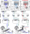
Anatomical changes in the somatosensory system after large sensory loss predict strategies to promote functional recovery after spinal cord injury
- PMID: 27212917
- PMCID: PMC4870913
- DOI: 10.4103/1673-5374.180741
Anatomical changes in the somatosensory system after large sensory loss predict strategies to promote functional recovery after spinal cord injury
Figures

Similar articles
-
Numb rats walk - a behavioural and fMRI comparison of mild and moderate spinal cord injury.Eur J Neurosci. 2003 Dec;18(11):3061-8. doi: 10.1111/j.1460-9568.2003.03062.x. Eur J Neurosci. 2003. PMID: 14656301
-
Time course of functional changes in locomotor and sensory systems after spinal cord lesions in lamprey.J Neurophysiol. 2019 Jun 1;121(6):2323-2335. doi: 10.1152/jn.00120.2019. Epub 2019 Apr 24. J Neurophysiol. 2019. PMID: 31017839
-
Somatosensory evoked potential changes and decompression timing for spinal cord function recovery and evoked potentials in rats with spinal cord injury.Brain Res Bull. 2019 Mar;146:7-11. doi: 10.1016/j.brainresbull.2018.12.003. Epub 2018 Dec 11. Brain Res Bull. 2019. PMID: 30550848
-
Intervention strategies to enhance anatomical plasticity and recovery of function after spinal cord injury.Adv Neurol. 1997;72:257-75. Adv Neurol. 1997. PMID: 8993704 Review.
-
Afferent input and sensory function after human spinal cord injury.J Neurophysiol. 2018 Jan 1;119(1):134-144. doi: 10.1152/jn.00354.2017. Epub 2017 Jul 12. J Neurophysiol. 2018. PMID: 28701541 Free PMC article. Review.
References
-
- Burton H, Fabri M. Ipsilateral intracortical connections of physiologically defined cutaneous representations in areas 3b and 1 of macaque monkeys: projections in the vicinity of the central sulcus. J Comp Neurol. 1995;355:508–538. - PubMed
-
- Kambi N, Halder P, Rajan R, Arora V, Chand P, Arora M, Jain N. Large-scale reorganization of the somatosensory cortex following spinal cord injuries is due to brainstem plasticity. Nat Commun. 2014;5:3602. - PubMed
Grants and funding
LinkOut - more resources
Full Text Sources
Other Literature Sources

