

Nerve growth factor promotes in vitro proliferation of neural stem cells from tree shrews
- PMID: 27212919
- PMCID: PMC4870915
- DOI: 10.4103/1673-5374.180743
Nerve growth factor promotes in vitro proliferation of neural stem cells from tree shrews
Abstract
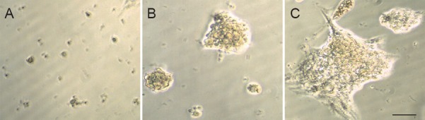
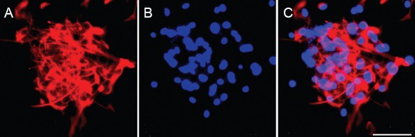
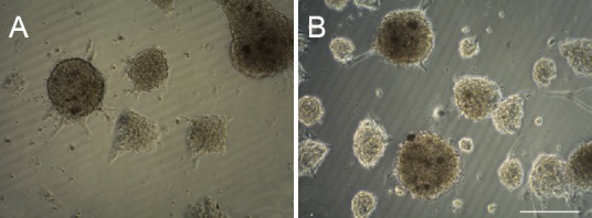
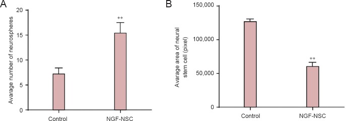
Neural stem cells promote neuronal regeneration and repair of brain tissue after injury, but have limited resources and proliferative ability in vivo. We hypothesized that nerve growth factor would promote in vitro proliferation of neural stem cells derived from the tree shrews, a primate-like mammal that has been proposed as an alternative to primates in biomedical translational research. We cultured neural stem cells from the hippocampus of tree shrews at embryonic day 38, and added nerve growth factor (100 μg/L) to the culture medium. Neural stem cells from the hippocampus of tree shrews cultured without nerve growth factor were used as controls. After 3 days, fluorescence microscopy after DAPI and nestin staining revealed that the number of neurospheres and DAPI/nestin-positive cells was markedly greater in the nerve growth factor-treated cells than in control cells. These findings demonstrate that nerve growth factor promotes the proliferation of neural stem cells derived from tree shrews.
Keywords: cell number; cell proliferation; cell therapy; embryo; hippocampus; in vitro; nerve growth factor; nerve regeneration; neural regeneration; neural stem cells; neurosphere; tree shrews.
Conflict of interest statement
Figures
References
-
- Aleksandrova MA, Podgornyi OV, Marei MV, Poltavtseva RA, Tsitrin EB, Gulyaev DV, Cherkasova LV, Revishchin AV, Korochkin LI, Khrushchov NG, and Sukhikh GN. Characteristics of human neural stem cells in vitro and after transplantation into rat brain. Bull Exp Biol Med. 2005;139:114–120. - PubMed
-
- Anderson DJ, Michelsohn A. Role of glucocorticoids in the chromaffin-neuron developmental decision. Int J Dev Neurosci. 1989;7:475–487. - PubMed
-
- Benoit BO, Savarese T, Joly M, Engstrom CM, Pang L, Reilly J, Recht LD, Ross AH, Quesenberry PJ. Neurotrophin channeling of neural progenitor cell differentiation. J Neurobiol. 2011;46:265–280. - PubMed
-
- Carito V, Nicolò S, Fiore M, Maccarone M, Tirassa P. Ocular nerve growth factor administration (oNGF) affects disease severity and inflammatory response in the brain of rats with experimental allergic encephalitis (EAE) Can J Physiol Pharmacol. 2015;29:1–8. - PubMed
-
- Chen MM, Zhao GW, He P, Jiang ZL, Xi X, Xu SH, Ma DM, Wang Y, Li YC, Wang GH. Improvement in the neural stem cell proliferation in rats treated with modified “Shengyu” decoction may contribute to the neurorestoration. J Ethnopharmacol. 2015;165:9–19. - PubMed
LinkOut - more resources
Full Text Sources
Other Literature Sources
