

Electroacupuncture at Baihui (DU20) acupoint up-regulates mRNA expression of NeuroD molecules in the brains of newborn rats suffering in utero fetal distress
- PMID: 27212921
- PMCID: PMC4870917
- DOI: 10.4103/1673-5374.180745
Electroacupuncture at Baihui (DU20) acupoint up-regulates mRNA expression of NeuroD molecules in the brains of newborn rats suffering in utero fetal distress
Abstract
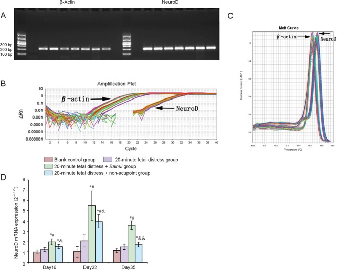
NeuroD plays a key regulatory effect on differentiation of neural stem cells into mature neurons in the brain. Thus, we assumed that electroacupuncture at Baihui (DU20) acupoint in newborn rats exposed to in utero fetal distress would influence expression of NeuroD. Electroacupuncture at Baihui was performed for 20 minutes on 3-day-old (Day 3) newborn Sprague-Dawley rats exposed to in utero fetal distress; electroacupuncture parameters consisted of sparse and dense waves at a frequency of 2-10 Hz. Real-time fluorescent quantitative PCR results demonstrated that mRNA expression of NeuroD, a molecule that indicates NeuroD, increased with prolonged time in brains of newborn rats, and peaked on Day 22. The level of mRNA expression was similar between Day 16 and Day 35. These findings suggest that electro acupuncture at Baihui acupoint could effectively increase mRNA expression of molecules involved in NeuroD in the brains of newborn rats exposed to in utero fetal distress.
Keywords: Baihui (DU20) acupoint; NeuroD; brain injury; electroacupuncture; hypoxic-ischemic brain injury; in utero fetal distress; nerve regeneration; nerve repair; neural regeneration; non-acupoint; real-time fluorescent quantitative PCR.
Conflict of interest statement
Figures
Similar articles
-
Electroacupuncture at the Baihui acupoint alleviates cognitive impairment and exerts neuroprotective effects by modulating the expression and processing of brain-derived neurotrophic factor in APP/PS1 transgenic mice.Mol Med Rep. 2016 Feb;13(2):1611-7. doi: 10.3892/mmr.2015.4751. Epub 2015 Dec 31. Mol Med Rep. 2016. PMID: 26739187 Free PMC article.
-
[Effect of electric acupuncture on the expression of NgR in the cerebral cortex, the medulla oblongata, and the spinal cord of hypertensive rats after cerebral infarction].Zhongguo Zhong Xi Yi Jie He Za Zhi. 2014 Mar;34(3):334-41. Zhongguo Zhong Xi Yi Jie He Za Zhi. 2014. PMID: 24758087 Chinese.
-
Hippocampal gene expression in a rat model of depression after electroacupuncture at the Baihui and Yintang acupoints.Neural Regen Res. 2014 Jan 1;9(1):76-83. doi: 10.4103/1673-5374.125333. Neural Regen Res. 2014. PMID: 25206746 Free PMC article.
-
[Local adenosine A1 receptors of baihui acupoint mediate cerebral ischemia tolerance induced by Electroacupuncture].Zhonghua Yi Xue Za Zhi. 2013 Feb 19;93(7):537-40. Zhonghua Yi Xue Za Zhi. 2013. PMID: 23660326 Chinese.
-
Electroacupuncture for control of blood glucose in diabetes: literature review.J Acupunct Meridian Stud. 2012 Feb;5(1):1-10. doi: 10.1016/j.jams.2011.11.018. Epub 2012 Jan 2. J Acupunct Meridian Stud. 2012. PMID: 22309901 Review.
Cited by
-
Efficacy of electro-acupuncture in postpartum with diastasis recti abdominis: A randomized controlled clinical trial.Front Public Health. 2022 Nov 15;10:1003361. doi: 10.3389/fpubh.2022.1003361. eCollection 2022. Front Public Health. 2022. PMID: 36483239 Free PMC article. Clinical Trial.
References
-
- Bjelke B, Andersson K, Ogren SO, Bolme P. Asphyctic lesion: proliferation of tyrosine hydroxylase-immunoreactive nerve cell bodies in the rat substantia nigra and functional changes in dopamine neurotransmission. Brain Res. 1991;543:1–9. - PubMed
-
- Chung JH, Lee EY, Jang MH, Kim CJ, Kim J, Ha E, Park HK, Choi S, Lee H, Park SH, Leem KH, Kim EH. Acupuncture decreases ischemia-induced apoptosis and cell proliferation in dentate gyrus of gerbils. Neuro Res. 2007;29:23–27. - PubMed
-
- Daval JL, Pourie G, Gean S, Lièvre V, Strazielle C, Blaise S, Vert P. Neonatal hypoxia triggers transient apoptosis followed by neurogenesis in the rat CA1 hippocampus. Pediatr Res. 2004;55:561–567. - PubMed
-
- Ehninqer D. Kempermann G Neurogenesis in the adult hippocampus. Cell Tissue Res. 2008;331:243–250. - PubMed
LinkOut - more resources
Full Text Sources
Other Literature Sources
