

Human umbilical cord blood-derived stem cells and brain-derived neurotrophic factor protect injured optic nerve: viscoelasticity characterization
- PMID: 27212930
- PMCID: PMC4870926
- DOI: 10.4103/1673-5374.180753
Human umbilical cord blood-derived stem cells and brain-derived neurotrophic factor protect injured optic nerve: viscoelasticity characterization
Abstract
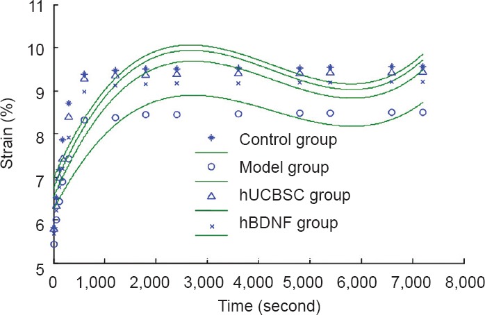
The optic nerve is a viscoelastic solid-like biomaterial. Its normal stress relaxation and creep properties enable the nerve to resist constant strain and protect it from injury. We hypothesized that stress relaxation and creep properties of the optic nerve change after injury. More-over, human brain-derived neurotrophic factor or umbilical cord blood-derived stem cells may restore these changes to normal. To validate this hypothesis, a rabbit model of optic nerve injury was established using a clamp approach. At 7 days after injury, the vitreous body re-ceived a one-time injection of 50 μg human brain-derived neurotrophic factor or 1 × 10(6) human umbilical cord blood-derived stem cells. At 30 days after injury, stress relaxation and creep properties of the optic nerve that received treatment had recovered greatly, with patho-logical changes in the injured optic nerve also noticeably improved. These results suggest that human brain-derived neurotrophic factor or umbilical cord blood-derived stem cell intervention promotes viscoelasticity recovery of injured optic nerves, and thereby contributes to nerve recovery.
Keywords: brain-derived neurotrophic factors; creep; histomorphology; human umbilical cord blood-derived stem cells; nerve regeneration; neural regeneration; optic nerve injury; stress relaxation; viscoelasticity.
Conflict of interest statement
Figures


Similar articles
-
Human umbilical cord blood stem cells and brain-derived neurotrophic factor for optic nerve injury: a biomechanical evaluation.Neural Regen Res. 2015 Jul;10(7):1134-8. doi: 10.4103/1673-5374.160110. Neural Regen Res. 2015. PMID: 26330839 Free PMC article.
-
Human umbilical cord blood-derived mesenchymal stem cells promote regeneration of crush-injured rat sciatic nerves.Neural Regen Res. 2012 Sep 15;7(26):2018-27. doi: 10.3969/j.issn.1673-5374.2012.26.003. Neural Regen Res. 2012. PMID: 25624833 Free PMC article.
-
Human amniotic epithelial cell transplantation for the repair of injured brachial plexus nerve: evaluation of nerve viscoelastic properties.Neural Regen Res. 2015 Feb;10(2):260-5. doi: 10.4103/1673-5374.152380. Neural Regen Res. 2015. PMID: 25883625 Free PMC article.
-
Functional recovery in acute traumatic spinal cord injury after transplantation of human umbilical cord mesenchymal stem cells.Crit Care Med. 2010 Nov;38(11):2181-9. doi: 10.1097/CCM.0b013e3181f17c0e. Crit Care Med. 2010. PMID: 20711072
-
Neurotrophic factors for spinal cord repair: Which, where, how and when to apply, and for what period of time?Brain Res. 2015 Sep 4;1619:36-71. doi: 10.1016/j.brainres.2014.10.049. Epub 2014 Nov 1. Brain Res. 2015. PMID: 25451132 Review.
Cited by
-
Unrestricted Somatic Stem Cells Loaded in Nanofibrous Conduit as Potential Candidate for Sciatic Nerve Regeneration.J Mol Neurosci. 2019 Jan;67(1):48-61. doi: 10.1007/s12031-018-1209-9. Epub 2018 Nov 27. J Mol Neurosci. 2019. PMID: 30484060
-
Roles of growth factors in eye development and ophthalmic diseases.Zhejiang Da Xue Xue Bao Yi Xue Ban. 2022 Nov 25;51(5):613-625. doi: 10.3724/zdxbyxb-2022-0603. Zhejiang Da Xue Xue Bao Yi Xue Ban. 2022. PMID: 36581579 Free PMC article. Review. English.
-
Neural stem cells over-expressing brain-derived neurotrophic factor promote neuronal survival and cytoskeletal protein expression in traumatic brain injury sites.Neural Regen Res. 2017 Mar;12(3):433-439. doi: 10.4103/1673-5374.202947. Neural Regen Res. 2017. PMID: 28469658 Free PMC article.
-
Umbilical Cord Blood and Serum for the Treatment of Ocular Diseases: A Comprehensive Review.Ophthalmol Ther. 2020 Jun;9(2):235-248. doi: 10.1007/s40123-020-00239-9. Epub 2020 Feb 27. Ophthalmol Ther. 2020. PMID: 32107737 Free PMC article. Review.
References
-
- Bi YY, Feng DF, Pan DC. Stem/progenitor cells: a potential source of retina-specific cells for retinal repair. Neurosci Res. 2009;65:215–221. - PubMed
-
- Bodanapally UK, Kathirkamanathan S, Geraymovych E, Mirvis SE, Choi AY, McMillan AB, Zhuo J, Shin RK. Diagnosis of traumatic optic neuropathy: application of diffusion tensor magnetic resonance imaging. J Neuroophthalmol. 2013;33:128–133. - PubMed
-
- Chakraborty SK, Banu LA, Rahman MF, Paul S. Cord blood stem cells - a dream for future medicine. Mymensingh Med J. 2014;23:614–620. - PubMed
LinkOut - more resources
Full Text Sources
Other Literature Sources
