. 2016:2016:6281376.
doi: 10.1155/2016/6281376. Epub 2016 Apr 24.

Flavonoids Extraction from Propolis Attenuates Pathological Cardiac Hypertrophy through PI3K/AKT Signaling Pathway

Affiliations

Flavonoids Extraction from Propolis Attenuates Pathological Cardiac Hypertrophy through PI3K/AKT Signaling Pathway

Guang-Wei Sun et al. Evid Based Complement Alternat Med. 2016.

Abstract

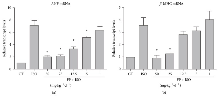
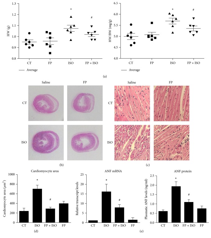
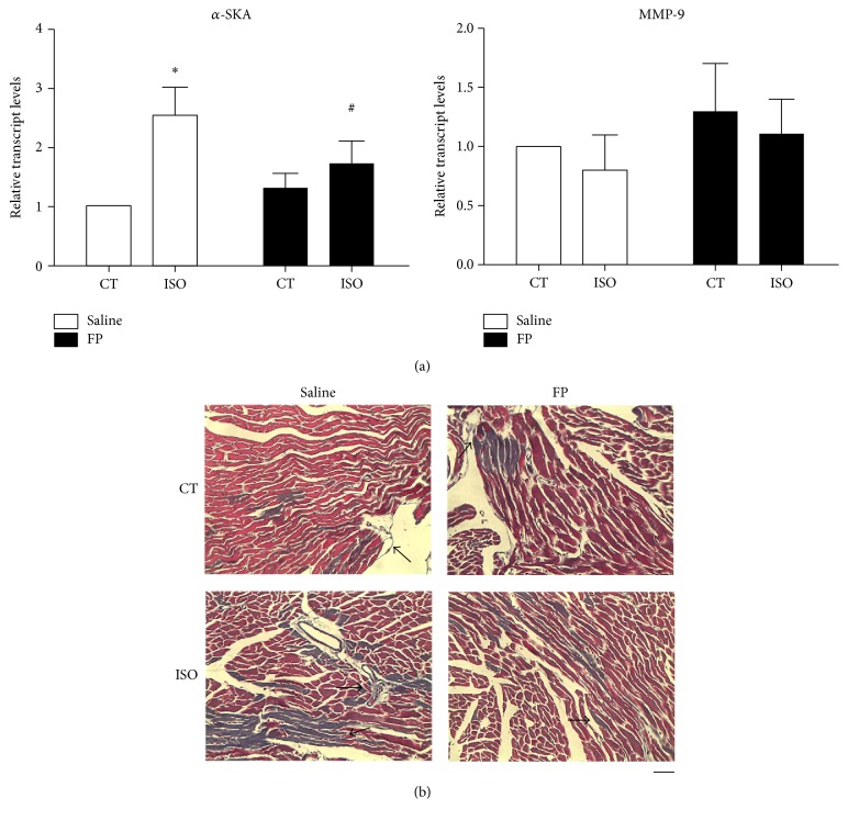
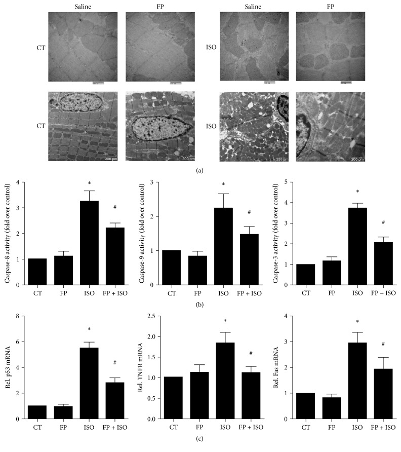
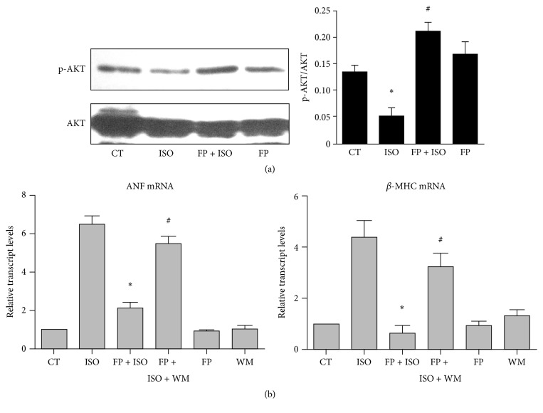
Propolis, a traditional medicine, has been widely used for a thousand years as an anti-inflammatory and antioxidant drug. The flavonoid fraction is the main active component of propolis, which possesses a wide range of biological activities, including activities related to heart disease. However, the role of the flavonoids extraction from propolis (FP) in heart disease remains unknown. This study shows that FP could attenuate ISO-induced pathological cardiac hypertrophy (PCH) and heart failure in mice. The effect of the two fetal cardiac genes, atrial natriuretic factor (ANF) and β-myosin heavy chain (β-MHC), on PCH was reversed by FP. Echocardiography analysis revealed cardiac ventricular dilation and contractile dysfunction in ISO-treated mice. This finding is consistent with the increased heart weight and cardiac ANF protein levels, massive replacement fibrosis, and myocardial apoptosis. However, pretreatment of mice with FP could attenuate cardiac dysfunction and hypertrophy in vivo. Furthermore, the cardiac protection of FP was suppressed by the pan-PI3K inhibitor wortmannin. FP is a novel cardioprotective agent that can attenuate adverse cardiac dysfunction, hypertrophy, and associated disorder, such as fibrosis. The effects may be closely correlated with PI3K/AKT signaling. FP may be clinically used to inhibit PCH progression and heart failure.


Figures

Figure 1
Figure 1
LC-MS base peak chromatograms of FP. The peak numbers refer to Table 1. FP (0–120 min).
Figure 2
Figure 2
FP represses isoproterenol-induced fetal gene reactivation in a dose-dependent manner. Intragastric injection of various doses of FP (1–50 mg·kg−1·d−1) for 7 days was followed by continuous infusion with isoproterenol (ISO, 25 mg·kg−1·d−1) for 7 days to experimentally induce heart hypertrophy. Heart tissues were collected and assayed for ANF (a) and β-MHC expression (b). The results are expressed as the means ± SE; n = 10 mice per group ( p < 0.05 compared with isoproterenol group).
Figure 3
Figure 3
Cardiac hypertrophy is reduced in mice treated by FP. Intragastric injection of FP (25 mg·kg−1·d−1) for 7 days followed by continuous infusion with isoproterenol (ISO, 25 mg·kg−1·d−1) for 7 days was sued to experimentally induce heart hypertrophy. (a) Animals were euthanized and the hearts were removed for hypertrophic evaluation. A comparison of HW (g) and HW/BW ratios (mg/g) for the animals is shown. (b) Representative histological sections of hearts stained with H&E. (c) Magnified images of histological sections in (b) used to determine CM cross-sectional area. (d) Quantification of CM cross-sectional area in the left ventricular wall. (e) mRNA expression and plasma protein levels of the hypertrophy marker ANF. The results are expressed as the means ± SE; n = 10 mice per group ( p < 0.05 compared to the control group; # p < 0.05 compared to the isoproterenol group).
Figure 4
Figure 4
FP treatment reduced associated disorder, such as fibrosis. (a) mRNA expression levels of the hypertrophy marker a-SKA and the fibrosis marker matrix metalloproteinase-9 (MMP-9). (b) Determination of fibrosis in histological sections by Masson's trichrome staining. Scale bar, 50 μm. The arrows show fibrotic areas. The results are expressed as the means ± SE; n = 10 mice per group ( p < 0.05 compared with control group; # p < 0.05 compared with isoproterenol group).
Figure 5
Figure 5
Effects of isoproterenol and FP on apoptotic damage and apoptosis-related gene expression in heart tissues. (a) Transmission electron microscopy of heart tissues. (b) The activities of caspase-8, caspase-9, and caspase-3 were measured using a fluorometric assay and expressed as the fold change over the control. (c) mRNA levels of p53, TNFR1, and Fas were determined by real-time RT-PCR. The levels of mRNA were normalized to GAPDH. Relative mRNA levels are shown using arbitrary units, and the value of the control group (CT) is defined as 1. The results are expressed as the means ± SE; n = 10 mice per group ( p < 0.05 compared to the control group; # p < 0.05 compared to the isoproterenol group).
Figure 6
Figure 6
FP prevents isoproterenol-induced cardiac remodeling in vivo, which is correlated with PI3K/AKT signaling. (a-b) Intragastric injection of FP (25 mg·kg−1·d−1) or saline (CT) for 7 days was followed by continuous infusion with isoproterenol (ISO, 25 mg·kg−1·d−1) for 7 days to experimentally induce heart hypertrophy. The animals were euthanized and the hearts were removed for hypertrophic evaluation. (a) Representative Western blotting assays and quantitative analysis of phosphorylated AKT in the hearts by drug treatment ( p < 0.05 compared to the control group; # p < 0.05 compared to the isoproterenol group). (b) Effect of a selective PI3K antagonist, wortmannin (WM), on isoproterenol-induced gene reactivation. The heart tissues were collected and assayed for ANF and β-MHC expression. The results are expressed as the means ± SE; n = 10 mice per group ( p < 0.05 compared to the isoproterenol group; # p < 0.05 compared to the FP-isoproterenol cotreated group).

References

    1. Abel E. D., Doenst T. Mitochondrial adaptations to physiological vs. pathological cardiac hypertrophy. Cardiovascular Research. 2011;90(2):234–242. doi: 10.1093/cvr/cvr015. - DOI - PMC - PubMed
    1. Allwood M. A., Kinobe R. T., Ballantyne L., et al. Heme oxygenase-1 overexpression exacerbates heart failure with aging and pressure overload but is protective against isoproterenol-induced cardiomyopathy in mice. Cardiovascular Pathology. 2014;23(4):231–237. doi: 10.1016/j.carpath.2014.03.007. - DOI - PubMed
    1. Anderson M., Moore D., Larson D. F. Comparison of isoproterenol and dobutamine in the induction of cardiac hypertrophy and fibrosis. Perfusion. 2008;23(4):231–235. doi: 10.1177/0267659108100708. - DOI - PubMed
    1. Aoyagi T., Matsui T. Phosphoinositide-3 kinase signaling in cardiac hypertrophy and heart failure. Current Pharmaceutical Design. 2011;17(18):1818–1824. doi: 10.2174/138161211796390976. - DOI - PMC - PubMed
    1. Benjamin M. M., Smith R. L., Grayburn P. A. Ischemic and functional mitral regurgitation in heart failure: natural history and treatment. Current Cardiology Reports. 2014;16(8):p. 517. doi: 10.1007/s11886-014-0517-0. - DOI - PubMed

LinkOut - more resources