. 2016;1(5):e86252.
doi: 10.1172/jci.insight.86252. Epub 2016 Apr 21.

Origin and evolution of the T cell repertoire after posttransplantation cyclophosphamide

Affiliations

Origin and evolution of the T cell repertoire after posttransplantation cyclophosphamide

Christopher G Kanakry et al. JCI Insight. 2016.

Abstract

Posttransplantation cyclophosphamide (PTCy) effectively prevents graft-versus-host disease (GVHD), but its immunologic impact is poorly understood. We assessed lymphocyte reconstitution via flow cytometry (n = 74) and antigen receptor sequencing (n = 35) in recipients of myeloablative, HLA-matched allogeneic BM transplantation using PTCy. Recovering T cells were primarily phenotypically effector memory with lower T cell receptor β (TRB) repertoire diversity than input donor repertoires. Recovering B cells were predominantly naive with immunoglobulin heavy chain locus (IGH) repertoire diversity similar to donors. Numerical T cell reconstitution and TRB diversity were strongly associated with recipient cytomegalovirus seropositivity. Global similarity between input donor and recipient posttransplant repertoires was uniformly low at 1-2 months after transplant but increased over the balance of the first posttransplant year. Blood TRB repertoires at ≥3 months after transplant were often dominated by clones present in the donor blood/marrow memory CD8+ compartment. Limited overlap was observed between the TRB repertoires of T cells infiltrating the skin or gastrointestinal tract versus the blood. Although public TRB sequences associated with herpesvirus- or alloantigen-specific CD8+ T cells were detected in some patients, posttransplant TRB and IGH repertoires were unique to each individual. These data define the immune dynamics occurring after PTCy and establish a benchmark against which immune recovery after other transplantation approaches can be compared.


Figures

Figure 1
Figure 1. The association of clinical factors with lymphocyte recovery after myeloablative, HLA-matched alloBMT using PTCy as single-agent GVHD prophylaxis.
Numerical reconstitution of lymphocytes, CD3+ T cells, CD3+CD4+ T cells, CD3+CD8+ T cells, CD3CD56+ NK cells, and CD19+ B cells is shown for (A) all patients and donors and stratified by (B) recipient cytomegalovirus (CMV) serostatus, (C) the detection of peripheral blood CMV reactivation after transplant occurring by the specified time point, or (D) the diagnosis of acute graft-versus-host disease (GVHD) by the specified time point. Error bars represent the standard error. Donor data in B are stratified by donor CMV serostatus. Donor and pretransplant recipient data are shown in D for comparison purposes only.
Figure 2
Figure 2. Lymphocyte subset numerical reconstitution is significantly associated with recipient CMV seropositivity.
(A and B) Numerical reconstitution of naive (CCR7+CD45RA+), central memory (CCR7+CD45RA), effector memory (CCR7CD45RA), and terminally differentiated CD45RA+ effector memory (TEMRA, CCR7CD45RA+) subsets of (A) CD3+CD4+ and (B) CD3+CD8+ T cell subsets is shown stratified by recipient pretransplant cytomegalovirus (CMV) serostatus. (C) Numerical reconstitution of CD19+ B cell subsets (naive mature, CD27CD24intCD38int; transitional, CD27CD24hiCD28hi; and memory, CD27+IgDCD24+CD38–/int) is shown stratified by recipient CMV serostatus. Donor data in all parts are stratified by donor CMV serostatus.
Figure 3
Figure 3. TRB and IGH sequence diversity in patients at 1 year after transplantation.
Unsorted peripheral blood mononuclear cells were assessed by survey-level sequencing for TRB and/or IGH. (A and B) There is a high correlation between 2 measurements of TRB diversity, clonality and Gini coefficient, for (A) patients treated with PTCy as single-agent GVHD prophylaxis (n = 34) and their donors (n = 15) (Pearson’s correlation, r = 0.967), as well as for (B) patients treated with methotrexate and a calcineurin inhibitor (CNI) for GVHD prophylaxis (n = 11) (Pearson’s correlation, r = 0.973). (C) TRB clonality for all 45 patients (both PTCy-treated and CNI-treated) is more strongly associated with recipient CMV seropositivity and posttransplant CMV peripheral blood reactivation than with history of acute or chronic graft-versus-host disease (GVHD). (D) IGH clonality in PTCy-treated recipients (n = 26) and CNI-treated recipients (n = 8) is similar to that of donors (n = 14), irrespective of CMV seropositivity, CMV reactivation, or GVHD history. R±/D± indicates recipient (R) and donor (D) CMV seropositivity or seronegativity.
Figure 4
Figure 4. Posttransplant TRB repertoires are unique to unrelated recipients.
Shown are pairwise similarity, calculated using the Bhattacharyya coefficient, between (A) TRB and (B) IGH repertoires of donors on the day of allograft donation (upper right in A and B) or of recipients at 1 year after transplant (lower left in A and B).
Figure 5
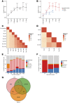
Figure 5. Posttransplant TRB repertoires become more similar to input donor repertoires over time during the first posttransplant year.
(A) Mean Bhattacharyya coefficients between the TRB repertoires of recipients at the indicated time points before or after transplant and the TRB repertoires of their related donors on the day of marrow donation. The number of donor/recipient pairs for whom a Bhattacharyya coefficient at each time point could be calculated is indicated. (B) Mean Bhattacharyya coefficients from A separated into 2 groups by donor CMV serostatus. The evolution toward a more “donor-like repertoire” over the first posttransplant year was more prominent in recipients of allografts from CMV-seropositive donors, in part due to the high frequency of CMV–specific memory T cells in both the donor and posttransplant recipient. (C) Pairwise TRB repertoire similarity matrix for donor and recipient 002-037. Both were CMV seropositive, and detectable CMV reactivation occurred in the recipient on day +41 after transplant. The donor repertoire was assessed on the day of allograft donation, and the recipient repertoire was assessed at the indicated time points before and after transplant. (D) Pairwise TRB repertoire similarity matrix for donor and recipient 002-031, who were both CMV seronegative. (E) Cumulative frequency of TRB sequences in the blood of recipient 002-037 on the indicated posttransplant days that could be presumptively mapped back to the donor’s peripheral blood (PB) repertoire. CD4+, CD8+, CD8+ naive, and CD8+ memory compartments were defined based on sorting of donor PB. The CD8+ CMV pp65-specific tetramer+ (HLA-A2– and HLA-B7–restricted) compartment was defined based on tetramer sorting of recipient samples at posttransplant days +83, +369, and +1,320. It was not possible to sort CMV pp65-specific tetramer+ cells from donor PB due to insufficient sample availability. (F) Cumulative frequency of TRB sequences in the blood of recipient 002-031 on the indicated posttransplant days that could be presumptively mapped back to the CD4+, CD8+, CD8+ naive, or CD8+ memory compartments of donor 002-031’s PB repertoire. (G) Venn diagram illustrating the overlap between the recipient CMV pp65 tetramer+ compartment and the TRB sequences that could be mapped back to the naive or memory compartment of donor 002-037, showing that nearly all CMV pp65 tetramer+ T cells that could be tracked were able to be traced to the memory compartment.
Figure 6
Figure 6. Dynamic repertoire changes occur after transplant.
Data from donor and recipient 001-006, both of whom were CMV seronegative, are shown. T cells were sorted from donor peripheral blood (PB) and BM samples by FACS and assessed by TRB sequencing. Unfractionated peripheral blood mononuclear cells collected from the recipient on posttransplant days +62, +188, and +363 were also assessed by TRB sequencing. (A) Absolute lymphocyte count (ALC) (top), clonality of the recipient TRB repertoire (middle), and similarity (Bhattacharyya coefficient) of the 001-006 recipient repertoire to the input donor repertoire (bottom) during the first posttransplant year. Posttransplant days of onset of grade I (1) and grade II (2) skin acute GVHD, start of immunosuppression with corticosteroids (3), and chronic GVHD (4) are indicated by the dotted vertical lines. (B) Relative frequency in donor 001-006’s PB and BM of TRB sequences that were observed in both physical compartments. The color of the point representing each sequence indicates the donor T cell phenotypic compartment in which that sequence was observed and from which it was sorted. (C and D) Left panels: Venn diagrams showing the overlap in unique TRB sequences between recipient 001-006 posttransplant PB, donor 001-006 PB, and donor 001-006 BM for (C) all TRB sequences or (D) the top first percentile of TRB sequences. The sequences observed in the 3 recipient posttransplant samples (days +62, +188, and +363) were grouped together for this analysis. Right panels: Relative frequency in the donor or in the recipient at the indicated time points of the TRB sequences that were observed in all 3 compartments (donor PB, donor BM, recipient PB). The color of the line representing each sequence indicates the donor T cell compartment (CD4+ naive, CD8+ naive, CD4+ memory, and CD8+ memory) in which that sequence was observed. TRB sequences that could not be unambiguously assigned to a specific donor T cell compartment are coded as “unassigned.” UD, undetected.
Figure 7
Figure 7. Overlap between TRB repertoires observed in blood or during evaluation for GVHD in the gastrointestinal tract and skin.
TRB sequencing was retrospectively performed on biopsies obtained from 4 recipients on this study, identified by the codes along the top of the figure, who underwent evaluation of both GI tract and skin for suspected GVHD. The posttransplant days on which GI and skin biopsies were obtained from each recipient are superimposed on the abdomen and chest, respectively, of the cutaway torso representing each patient. Recipients 001-007, 002-001, and 002-010 were female, but a male torso is displayed for all recipients for the sake of simplicity. The approximate location from which each GI tract biopsy was obtained is indicated by the blue dots; the location from which the skin biopsies (green dots) were obtained is not indicated. The days below the red dot superimposed on the vein in the right arm of each torso indicate the posttransplant dates of collection of blood used for TRB sequencing of unfractionated and/or sorted cells. The Venn diagram below each torso indicates the total number of unique TRB sequences collectively found in the blood samples (PBMC, red), skin biopsies (green), or GI biopsies (blue) obtained from each recipient, as well as the number of TRB sequences that were shared between the 3 compartments.
Figure 8
Figure 8. Tracking T cell clones from donor blood, BM, and tissue to recipient blood and tissue.
A CMV-seropositive patient (001-007) with AML received an allograft from her CMV-seronegative sister who had a history of inflammatory bowel disease. TRB sequencing was performed on donor blood and BM collected on the day of donation, and on tissue from gastrointestinal biopsies performed 8 months prior to donation that showed lymphocytic colitis. Three TRB sequences, denoted by the amino acid sequences encoded in their CDR3 regions, were shared between the BM, blood, and gastrointestinal biopsies from the donor and between the blood and gastrointestinal biopsies from the recipient. Although all 3 sequences were observed in the posttransplant blood and gastrointestinal tract biopsies of the recipient, who died of complications of gastrointestinal grade III acute GVHD, none were observed in a skin biopsy from the recipient, who also showed acute skin GVHD. Circles indicate the site of tissue sampling, and the color of the circle indicates the TRB sequence detected at that site.

References

    1. Kanakry CG, Fuchs EJ, Luznik L. Modern approaches to HLA-haploidentical blood or marrow transplantation. Nat Rev Clin Oncol. 2016;13(1):10–24. - PMC - PubMed
    1. Luznik L, et al. HLA-haploidentical bone marrow transplantation for hematologic malignancies using nonmyeloablative conditioning and high-dose, posttransplantation cyclophosphamide. Biol Blood Marrow Transplant. 2008;14(6):641–650. doi: 10.1016/j.bbmt.2008.03.005. - DOI - PMC - PubMed
    1. Luznik L, et al. High-dose cyclophosphamide as single-agent, short-course prophylaxis of graft-versus-host disease. Blood. 2010;115(16):3224–3230. doi: 10.1182/blood-2009-11-251595. - DOI - PMC - PubMed
    1. Bolanos-Meade J, et al. HLA-haploidentical bone marrow transplantation with posttransplant cyclophosphamide expands the donor pool for patients with sickle cell disease. Blood. 2012;120(22):4285–4291. doi: 10.1182/blood-2012-07-438408. - DOI - PMC - PubMed
    1. Bashey A, et al. T-cell-replete HLA-haploidentical hematopoietic transplantation for hematologic malignancies using post-transplantation cyclophosphamide results in outcomes equivalent to those of contemporaneous HLA-matched related and unrelated donor transplantation. J Clin Oncol. 2013;31(10):1310–1316. doi: 10.1200/JCO.2012.44.3523. - DOI - PubMed