. 2016 May 21;17(5):787.
doi: 10.3390/ijms17050787.

Functional and Biochemical Characterization of Three Recombinant Human Glucose-6-Phosphate Dehydrogenase Mutants: Zacatecas, Vanua-Lava and Viangchan

Affiliations

Functional and Biochemical Characterization of Three Recombinant Human Glucose-6-Phosphate Dehydrogenase Mutants: Zacatecas, Vanua-Lava and Viangchan

Saúl Gómez-Manzo et al. Int J Mol Sci. .

Abstract

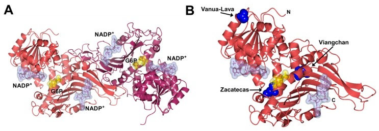
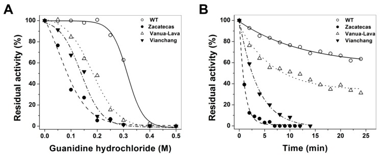
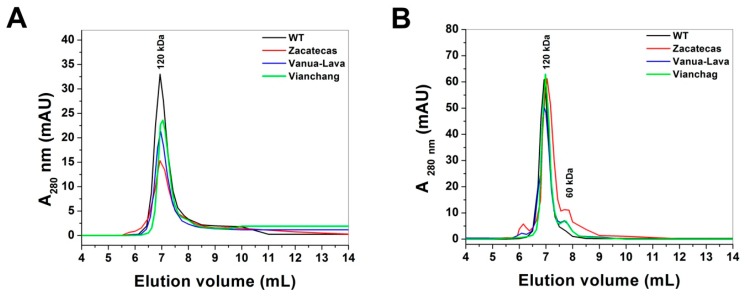
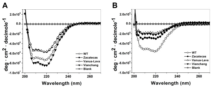
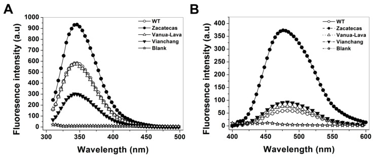
Glucose-6-phosphate dehydrogenase (G6PD) deficiency in humans causes severe disease, varying from mostly asymptomatic individuals to patients showing neonatal jaundice, acute hemolysis episodes or chronic nonspherocytic hemolytic anemia. In order to understand the effect of the mutations in G6PD gene function and its relation with G6PD deficiency severity, we report the construction, cloning and expression as well as the detailed kinetic and stability characterization of three purified clinical variants of G6PD that present in the Mexican population: G6PD Zacatecas (Class I), Vanua-Lava (Class II) and Viangchan (Class II). For all the G6PD mutants, we obtained low purification yield and altered kinetic parameters compared with Wild Type (WT). Our results show that the mutations, regardless of the distance from the active site where they are located, affect the catalytic properties and structural parameters and that these changes could be associated with the clinical presentation of the deficiency. Specifically, the structural characterization of the G6PD Zacatecas mutant suggests that the R257L mutation have a strong effect on the global stability of G6PD favoring an unstable active site. Using computational analysis, we offer a molecular explanation of the effects of these mutations on the active site.

Keywords: glucose-6-phosphate dehydrogenase (G6PD) deficiency; human G6PD mutants; steady state kinetics; structural characterization; thermostability.


Figures

Figure 1
Figure 1
Crystallographic structure of the human glucose-6-phosphate dehydrogenase (G6PD) enzyme: (A) The human Wild Type (WT) structure of G6PD dimer (PDB entries 2BHL and 2BH9) showing the NADP+ binding (ice blue molecular surface) at the structural and coenzyme sites and the G6P site (yellow molecular surface). The two monomers are shown in pale crimson and dark purple; (B) Zacatecas, Viangchan and Vanua-Lava mutants are shown as blue spheres in the human G6PD structure. The graphical representations were also constructed with CCP4mg version 2.10.4 [18].
Figure 2
Figure 2
Heterologous expression of glucose-6-Phosphate Dehydrogenases (G6PDs) in Escherichia coli BL21(DE3)Δzwf::kanr: (A) expression of human WT G6PD; (B) G6PD Zacatecas; and two Class II mutants; (C) G6PD Vanua-Lava and (D) G6PD Viangchan. Sonicated cell were centrifuged, and the resulting supernatants were used to measure the specific activity in each case. The G6PD specific activity was used as indicative of the expression levels of soluble recombinant protein. The standard deviations represent the value of triplicates samples.
Figure 3
Figure 3
Thermostability assays of recombinant Wild Type glucose-6-phosphate dehydrogenase (WT G6PD) and the three mutants with different NADP+ concentrations: (A) WT G6PD; (B) G6PD Zacatecas (R257L); (C) G6PD Vanua-Lava (L128P); and (D) G6PD Viangchan (V291M). In all cases, 200 ng of total protein was used. Residual activity was expressed as a percentage of the activity for the same sample incubated at 37 °C. The assays were performed in duplicate; standard errors were lower than 5%. NADP+ concentrations: without NADP+ (○), 10 µM (•), 100 µM (∆) and 500 µM (▼) NADP+, respectively.
Figure 4
Figure 4
The mutations decrease the thermal stability of WT G6PD enzyme. The determination of melting temperature (Tm) was monitored by recording the change in the CD signal at 222 nm when the temperature was increased progressively from 20 to 90 °C at 1°/2.5 min. In all cases, both the WT G6PD and the mutants were recorded at 0.8 mg/mL in 25 mM phosphate buffer pH 7.35. Experiments were performed in duplicate; standard errors were less than 5%.
Figure 5
Figure 5
Stability of glucose-6-phosphate dehydrogenases (G6PDs) incubated with different concentrations of Gdn-HCl and inactivation assays. (A) Effects of Gdn-HCl on the activity of WT G6PD and the G6PD Zacatecas, Vanua-Lava and Viangchan mutants. All the enzymes were incubated at 0.2 mg/mL in 50 mM phosphate buffer pH 7.35 in the presence of the indicated concentrations of Gdn-HCl for 2 h at 37 °C; (B) Inactivation of WT G6PD and the mutants by 0.25 M at 37 °C. At the indicated times, aliquots were withdrawn from the samples and assayed for residual activity. In both assays, residual activity was expressed as a percentage of the activity for the same sample measured at 25 °C without Gdn-HCl and all enzymes were prepared and diluted immediately before used. Both assays were performed in duplicate; standard errors were less than 5%.
Figure 6
Figure 6
Gel filtration chromatography to evaluate the protein stability of glucose-6-phosphate dehydrogenases (G6PDs). The stability of native G6PDs dimers without (A) or with (B) Gdn-HCl were tested by FPLC, incubating the enzymes (0.2 mg/mL) at 37 °C for 2 h and then loading them onto a size-exclusion chromatography column. Thirty microliters samples of G6PDs protein solutions were loaded on Shodex Protein® KW-802.5 column coupled to ÄKTA Primes FPLC system (Amersham Pharmacia Biotech, Piscataway, NJ, USA) previously equilibrated with 50 mM phosphate buffer at pH 7.35; flow rate: 1.0 mL/min.
Figure 7
Figure 7
Spectroscopic characterization of recombinant human G6PD enzymes. Far-UV CD spectra of WT G6PD and mutants without (A) or with (B) 0.25 M Gdn-HCl were performed in spectropolarimeter (Jasco J-810®) equipped with a Peltier thermostatted cell holder; standard errors were less than 4%. In all cases, the protein concentration was 0.2 mg/mL and incubated by 2 h at 37 °C and after than measured by CD. For both trials, the spectra of blanks were subtracted from those that contained the protein.
Figure 8
Figure 8
Conformational changes in the tertiary structure of recombinant human WT G6PD enzymes and mutants. (A) Fluorescence emission spectra were performed in a Perkin-Elmer LS-55 fluorescence spectrometer. The assays were conducted with a protein concentration of 0.1 mg/mL; (B) ANS fluorescence spectra were obtained using an excitation wavelength of 395 nm and recording emission spectra from 400 to 600 nm. Values obtained from buffer containing ANS without protein (open stars) were subtracted from the recordings with protein. The experimental conditions for all the experiments are described in the Materials and Methods Section.
Figure 9
Figure 9
Structural comparison between the human WT G6PD enzyme and the Class I G6PD Zacatecas mutant. (A) Crystallographic structure of human WT-G6PD enzyme (pale crimson) (PDB entry 2BH9); (B) Minimized model of the Class I G6PD Zacatecas variant (pale crimson) with the in silico R257L mutation. Note that the R257 (black cylinders) residue forms a weak cation–π interaction with W462 and a salt bridge with E473 in (A), and their absence in the in silico R257L mutant in (B). Distances are in Å; (C) Alignment of amino acid sequence of G6PD with homologs from different species from Gorilla gorilla gorilla (G3RMM2), Homo sapiens (P11413-3), Chlorocebus sabaeus (A0A0D9R328), Papio anubis (A9CB69), Macaca mulatta (H9ESV7), Macaca fascicularis (G7Q228), Equus caballus (F7DMG5), Camelus dromedarius (G1EHI3), Rhinolophus ferrumequinum (B2KIK5), Mustela putorius furo (M3YE89), Bos taurus (F1MMK2), Rattus norvegicus (P05370), Canis lupus familiaris (E2R0I9), Cavia porcellus (H0W6W1), Macropus robustus (Q29492), Sarcophilus harrisii (G3VHF4), Myotis brandtii (S7N6K2), Ornithorhynchus anatinus (F7DZC3), Sorex araneus (B3RFE2), Mus caroli (A0FF42), Canis lupus familiaris (J9P9E9), Crotalus adamanteus (A0A0F7Z7U2), Pelodiscus sinensis (K7FZ73), Bos indicus (Q7YS37), Xiphophorus maculatus (M4AS60), Lepisosteus oculatus (W5NAB4), Oreochromis niloticus (I3KK42), Ambystoma mexicanum (Q76BG5), Danio rerio (E7FDY7), Gasterosteus aculeatus (G3NFB2), Cephaloscyllium umbratile (Q76BC2), Callorhinchus mili (V9KLG6), Lepisosteus osseus (Q76BF1), Rhabdosargus sarba (Q4G339), Takifugu rubripes (H2UQV8), Scleropages formosus (A0A0P7V266), Xenopus tropicalis (F6XH10), Stegodyphus mimosarum (A0A087TRQ6), Capitella teleta (R7TN68), Ciona intestinalis (F7AX62), Branchiostoma floridae (C3YV81), Tribolium castaneum (D6WKK9), Rhipicephalus microplus (Q45R45), Lottia gigantea (V4AWI8), Triatoma infestans (A0A023F8E9), Anopheles gambiae (H2KMF3), Apis mellifera (A0A023FG14), Musca domestica (T1PD22), Aedes aegypti (Q0IEL8) performed with BioEdit V.7.2.5. The uniform colors indicate conserved amino acid in the sequences reported. Colorless represent non-conserved amino acid sequences. The arrows indicate the position R257 and E473 residues are highly conserved in different organisms.
Figure 9
Figure 9
Structural comparison between the human WT G6PD enzyme and the Class I G6PD Zacatecas mutant. (A) Crystallographic structure of human WT-G6PD enzyme (pale crimson) (PDB entry 2BH9); (B) Minimized model of the Class I G6PD Zacatecas variant (pale crimson) with the in silico R257L mutation. Note that the R257 (black cylinders) residue forms a weak cation–π interaction with W462 and a salt bridge with E473 in (A), and their absence in the in silico R257L mutant in (B). Distances are in Å; (C) Alignment of amino acid sequence of G6PD with homologs from different species from Gorilla gorilla gorilla (G3RMM2), Homo sapiens (P11413-3), Chlorocebus sabaeus (A0A0D9R328), Papio anubis (A9CB69), Macaca mulatta (H9ESV7), Macaca fascicularis (G7Q228), Equus caballus (F7DMG5), Camelus dromedarius (G1EHI3), Rhinolophus ferrumequinum (B2KIK5), Mustela putorius furo (M3YE89), Bos taurus (F1MMK2), Rattus norvegicus (P05370), Canis lupus familiaris (E2R0I9), Cavia porcellus (H0W6W1), Macropus robustus (Q29492), Sarcophilus harrisii (G3VHF4), Myotis brandtii (S7N6K2), Ornithorhynchus anatinus (F7DZC3), Sorex araneus (B3RFE2), Mus caroli (A0FF42), Canis lupus familiaris (J9P9E9), Crotalus adamanteus (A0A0F7Z7U2), Pelodiscus sinensis (K7FZ73), Bos indicus (Q7YS37), Xiphophorus maculatus (M4AS60), Lepisosteus oculatus (W5NAB4), Oreochromis niloticus (I3KK42), Ambystoma mexicanum (Q76BG5), Danio rerio (E7FDY7), Gasterosteus aculeatus (G3NFB2), Cephaloscyllium umbratile (Q76BC2), Callorhinchus mili (V9KLG6), Lepisosteus osseus (Q76BF1), Rhabdosargus sarba (Q4G339), Takifugu rubripes (H2UQV8), Scleropages formosus (A0A0P7V266), Xenopus tropicalis (F6XH10), Stegodyphus mimosarum (A0A087TRQ6), Capitella teleta (R7TN68), Ciona intestinalis (F7AX62), Branchiostoma floridae (C3YV81), Tribolium castaneum (D6WKK9), Rhipicephalus microplus (Q45R45), Lottia gigantea (V4AWI8), Triatoma infestans (A0A023F8E9), Anopheles gambiae (H2KMF3), Apis mellifera (A0A023FG14), Musca domestica (T1PD22), Aedes aegypti (Q0IEL8) performed with BioEdit V.7.2.5. The uniform colors indicate conserved amino acid in the sequences reported. Colorless represent non-conserved amino acid sequences. The arrows indicate the position R257 and E473 residues are highly conserved in different organisms.

Similar articles

Cited by

References

    1. Pai G.S., Sprenkle J.A., Do T.T., Mareni C.E., Migeon B.R. Localization of loci for hypoxanthine phosphoribosyltransferase and glucose-6-phosphate dehydrogenase and biochemical evidence of nonrandom X chromosome expression from studies of a human X-autosome translocation. Proc. Natl. Acad. Sci. USA. 1980;77:2810–2813. doi: 10.1073/pnas.77.5.2810. - DOI - PMC - PubMed
    1. Mason P.J., Bautista J.M., Gilsanz F. G6PD deficiency: The genotype-phenotype association. Blood Rev. 2007;5:267–283. doi: 10.1016/j.blre.2007.05.002. - DOI - PubMed
    1. Nkhoma E.T., Poole C., Vannappagari V., Hall S.A., Beutler E. The global prevalence of glucose-6-phosphate dehydrogenase deficiency: A systematic review and meta-analysis. Blood Cells Mol. Dis. 2009;42:267–278. doi: 10.1016/j.bcmd.2008.12.005. - DOI - PubMed
    1. Beutler E. G6PD deficiency. Blood. 1994;84:3613–3636. - PubMed
    1. Cappellini D., Fiorelli G. Glucose-6-phosphate dehydrogenase deficiency. Lancet. 2008;606:64–74. doi: 10.1016/S0140-6736(08)60073-2. - DOI - PubMed

Substances

LinkOut - more resources