. 2016 May 19;8(5):138.
doi: 10.3390/v8050138.

Pediatric and Adult High-Grade Glioma Stem Cell Culture Models Are Permissive to Lytic Infection with Parvovirus H-1

Affiliations

Pediatric and Adult High-Grade Glioma Stem Cell Culture Models Are Permissive to Lytic Infection with Parvovirus H-1

Rafael Josupeit et al. Viruses. .

Abstract

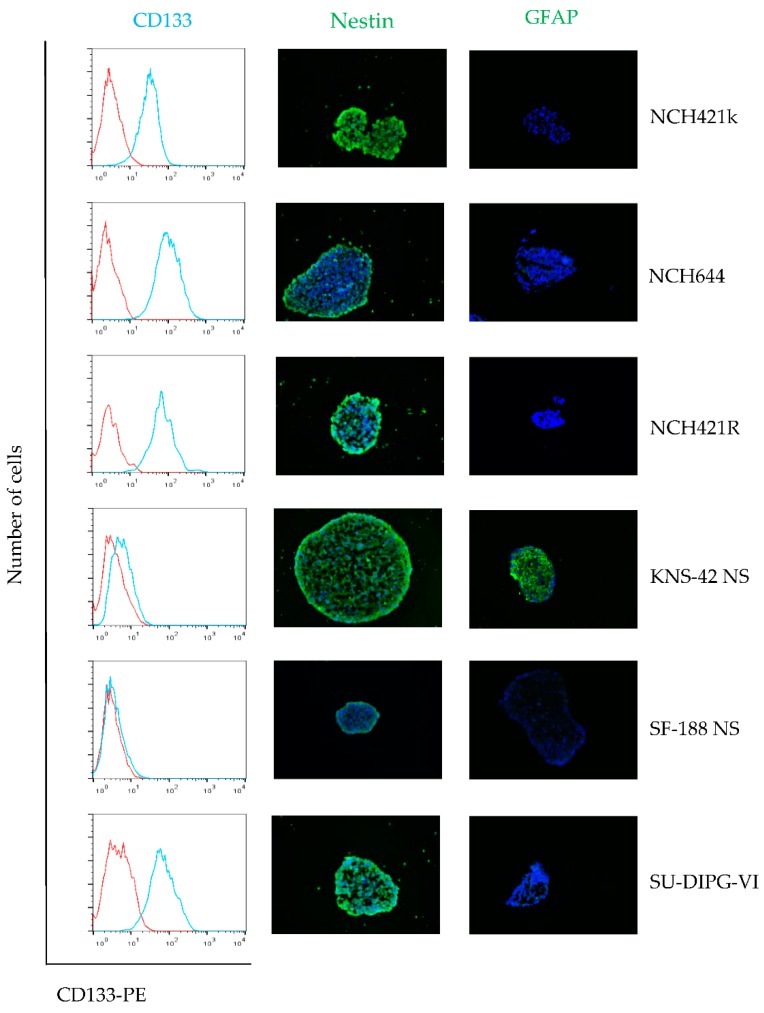
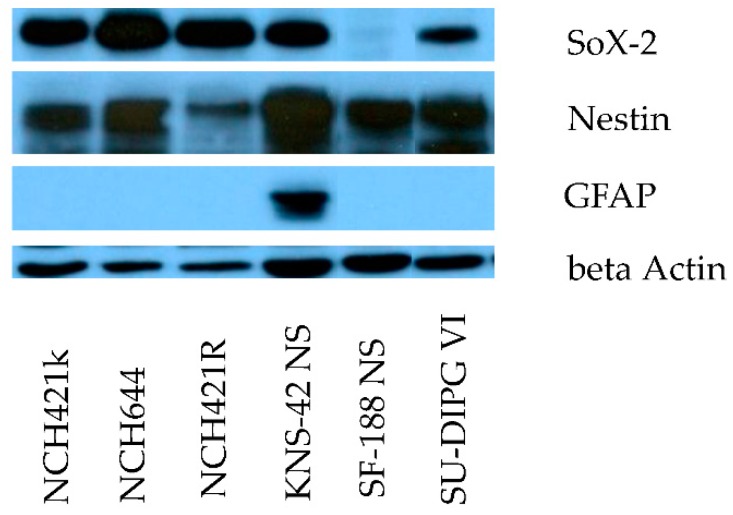
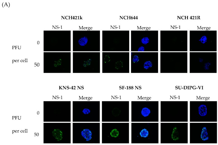
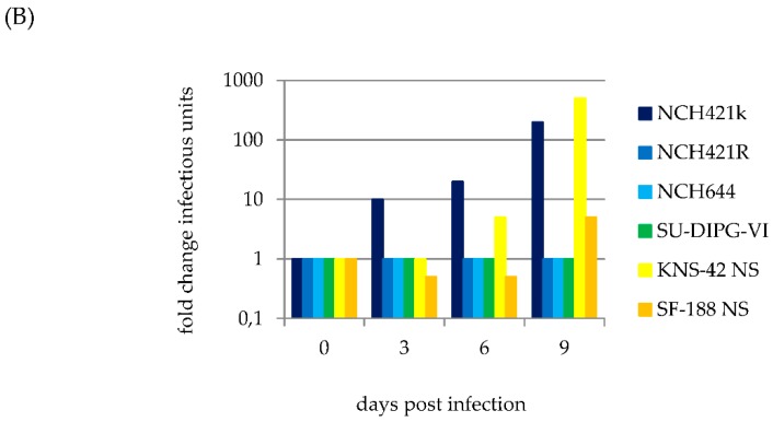
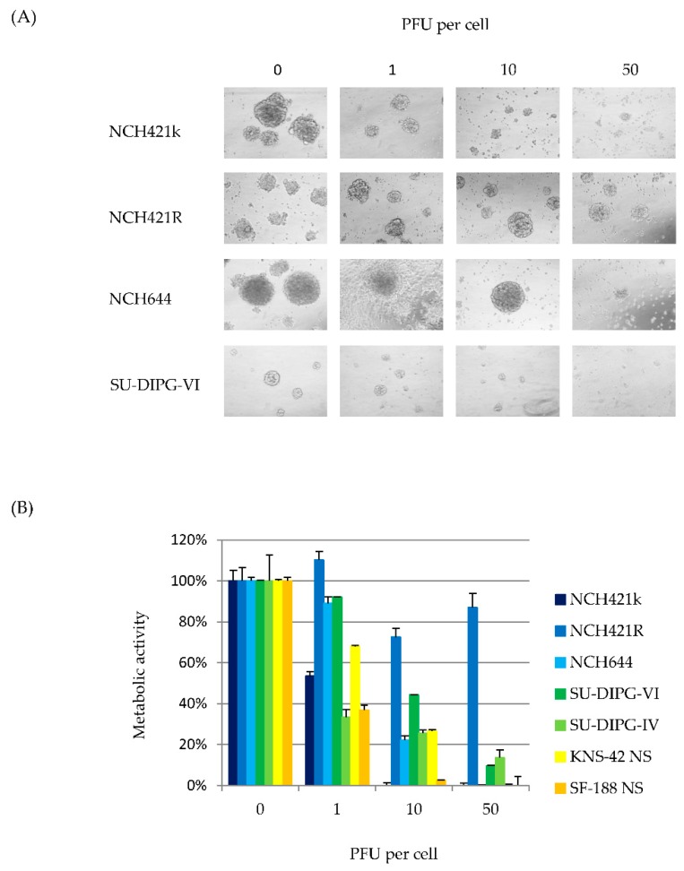
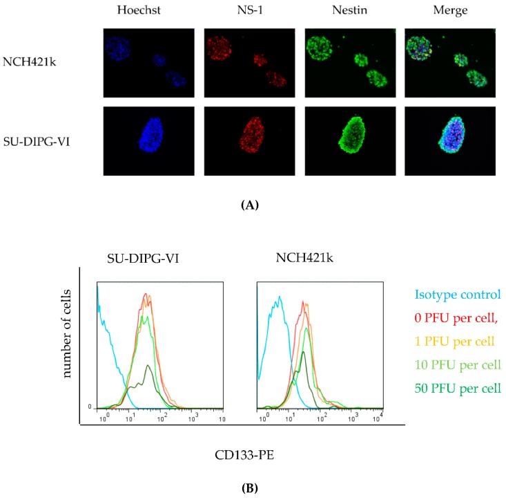
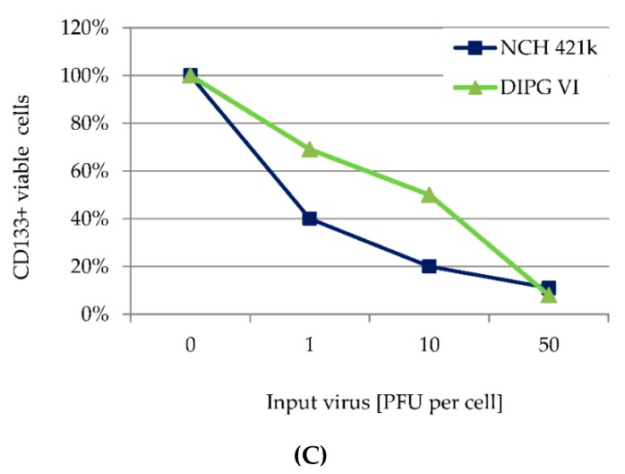
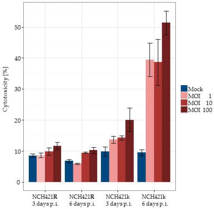
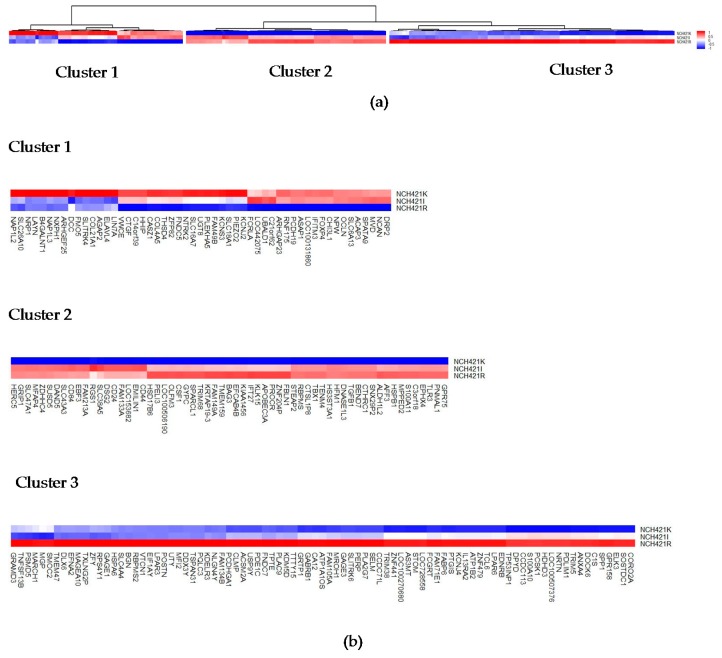
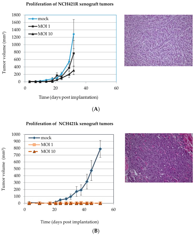
Combining virus-induced cytotoxic and immunotherapeutic effects, oncolytic virotherapy represents a promising therapeutic approach for high-grade glioma (HGG). A clinical trial has recently provided evidence for the clinical safety of the oncolytic parvovirus H-1 (H-1PV) in adult glioblastoma relapse patients. The present study assesses the efficacy of H-1PV in eliminating HGG initiating cells. H-1PV was able to enter and to transduce all HGG neurosphere culture models (n = 6), including cultures derived from adult glioblastoma, pediatric glioblastoma, and diffuse intrinsic pontine glioma. Cytotoxic effects induced by the virus have been observed in all HGG neurospheres at half maximal inhibitory concentration (IC50) doses of input virus between 1 and 10 plaque forming units per cell. H-1PV infection at this dose range was able to prevent tumorigenicity of NCH421k glioblastoma multiforme (GBM) "stem-like" cells in NOD/SCID mice. Interestingly NCH421R, an isogenic subclone with equal capacity of xenograft formation, but resistant to H-1PV infection could be isolated from the parental NCH421k culture. To reveal changes in gene expression associated with H-1PV resistance we performed a comparative gene expression analysis in these subclones. Several dysregulated genes encoding receptor proteins, endocytosis factors or regulators innate antiviral responses were identified and represent intriguing candidates for to further study molecular mechanisms of H-1PV resistance.

Keywords: DIPG; H-1PV; glioma stem-like cells; high-grade glioma; oncolytic virus; parvovirus H-1; pediatric glioblastoma; tumor initiating cells.


Figures

Figure 1
Figure 1
High-grade glioma (HGG) neurosphere cultures show stem cell features. Expression of neuroepithelial stem cell marker proteins CD133 and Nestin, and the astrocyte lineage marker glial fibrillary acidic protein (GFAP) in neurosphere cultures were determined by fluorescence-activated cell sorting (FACS) analysis and immunofluorescence microscopy. CD133 expression was assayed by FACS (cyan) and isotype control is depicted as the (red) respectively. Immunofluorescence for Nestin (green) and GFAP (green) merged with Hoechst 33342 (blue), magnification 400×.
Figure 2
Figure 2
Stem-like cell expression pattern in HGG neurosphere culture models. Expression of neuroepithelial stem cell marker proteins SOX-2 and Nestin, and the astrocyte lineage marker GFAP in neurosphere cultures by Western blot. Detection of beta actin served as loading control.
Figure 3
Figure 3
H-1 parvovirus (H-1PV) initiates replication in adult and pediatric HGG neurospheres. Indicated HGG neurosphere cultures were infected with H-1PV (one PFU per cell) six days post seeding. (A) Three days post infection, the initiation of virus replication was measured by fluorescence microscopy after cell immuno-staining for the nonstructural viral protein 1 (NS1) (green), co-staining of neurospheres with Hoechst 33342 (blue) is shown in merge panels; (B) H-1PV virus production was assayed in a time course experiment by quantification of infectious particles as described in Section 2.5.
Figure 3
Figure 3
H-1 parvovirus (H-1PV) initiates replication in adult and pediatric HGG neurospheres. Indicated HGG neurosphere cultures were infected with H-1PV (one PFU per cell) six days post seeding. (A) Three days post infection, the initiation of virus replication was measured by fluorescence microscopy after cell immuno-staining for the nonstructural viral protein 1 (NS1) (green), co-staining of neurospheres with Hoechst 33342 (blue) is shown in merge panels; (B) H-1PV virus production was assayed in a time course experiment by quantification of infectious particles as described in Section 2.5.
Figure 4
Figure 4
H-1PV induces cytotoxic effects in HGG “stem-like” cells. (A) Microscopic images of neurosphere cultures infected with indicated doses of H-1PV, nine days post infection, 400× magnification. Neurosphere morphology was determined prior to the toxicity assay; (B) At day nine after H-1PV infection at indicated multiplicities, cell metabolic activity was determined by WST-1 assay. Average values with error bars (SEM) from six independent experiments are shown. In all cultures but the resistant subclone NCH421R, 50% inhibition (LD50) was reached after infection with one to ten PFU per cell. After H-1PV infection decrease in metabolic activity in all cell cultures as compared to NCH421R was highly significant (p ≤ 0.001).
Figure 5
Figure 5
H-1V infection is cytotoxic to Nestin and CD133 positive cells. (A) Immunofluorescence of two HGG neurosphere cell lines infected with one PFU per cell nine days post seeding, which corresponds to three days post infection. Nuclei of the cells were stained with Hoechst 33342 (blue), NS-1 (red), Nestin (green) and images were merged, 400× magnification. Foci of NS-1 expression colocalize to nuclear staining with Hoechst in Nestin positive cells; (B) Cytotoxic effects of H-1PV on CD133-positive cells were assessed by FACS determination of the virus dose-dependent induction of CD133+ cell number at day three after infection. The isotype control was performed using uninfected cells (cyan); (C) 7AAD negative, i.e., viable cells of two HGG neurosphere cell cultures as determined by FACS nine days post infection with H-1PV at the MOIs indicated.
Figure 5
Figure 5
H-1V infection is cytotoxic to Nestin and CD133 positive cells. (A) Immunofluorescence of two HGG neurosphere cell lines infected with one PFU per cell nine days post seeding, which corresponds to three days post infection. Nuclei of the cells were stained with Hoechst 33342 (blue), NS-1 (red), Nestin (green) and images were merged, 400× magnification. Foci of NS-1 expression colocalize to nuclear staining with Hoechst in Nestin positive cells; (B) Cytotoxic effects of H-1PV on CD133-positive cells were assessed by FACS determination of the virus dose-dependent induction of CD133+ cell number at day three after infection. The isotype control was performed using uninfected cells (cyan); (C) 7AAD negative, i.e., viable cells of two HGG neurosphere cell cultures as determined by FACS nine days post infection with H-1PV at the MOIs indicated.
Figure 6
Figure 6
H-1PV infection induces lytic infection of HGG neurospheres in vitro. Lactate dehydrogenase (LDH)-release assays were performed on NCH421k and NCH421R cells at days 3 and 6 after infection with H-1PV at increasing MOIs. Specific lysis is given in relation to completely lysed cells by detergent treatment. Means from six replicates and respective standard errors are displayed.
Figure 7
Figure 7
Comparative transcriptome analysis of the NCH421k and its partially and fully H-1PV resistant subclones NCH421I and NCH421R. (a) Unsupervised clustering of the 201 genes most differentially expressed between NCH421k, NCH421I and the resistant subclone NCH421R. Quantitative comparison of the three profiles reveals genes that are up (red) or down (blue) regulated in resistant NCH421R cells in comparison with NCH421I and NCH421k cells showing high susceptibility to H-1PV infection; (b) Each of the three main clusters is shown independently in order to allow a more detailed analysis.
Figure 8
Figure 8
H-1PV infection suppresses tumorigenicity of HGG neurospheres in vivo. (A) NCH421R and (B) NCH421k neurosphere cultures were infected with H-1PV (MOI = one or ten PFU/cell) 4 h prior to subcutaneous implantation into the right flank of NOD/SCID mice (106 cells per animal). Mock-treated NCH421k cells served as non-treatment controls. Mock-infected NCH421k and NCH421R xenografts were confirmed to preserve a HGG histomorphology (hematoxylin/eosin, 400× magnification). Significant differences in xenograft tumor proliferation between mock infected cells and H-1PV infected cells were only observed in NCH421k xenograft bearing animals (p < 0.001).

Similar articles

Cited by

References

    1. Sturm D., Bender S., Jones D.T., Lichter P., Grill J., Becher O., Hawkins C., Majewski J., Jones C., Costello J.F., et al. Paediatric and adult glioblastoma: multiform (epi)genomic culprits emerge. Nat. Rev. Cancer. 2014;14:92–107. doi: 10.1038/nrc3655. - DOI - PMC - PubMed
    1. Fangusaro J., Warren K.E. Unclear standard of care for pediatric high grade glioma patients. J. Neurooncol. 2013;113:341–342. doi: 10.1007/s11060-013-1104-8. - DOI - PMC - PubMed
    1. Hardesty D.A., Sanai N. The value of glioma extent of resection in the modern neurosurgical era. Front. Neurol. 2012;3:140. doi: 10.3389/fneur.2012.00140. - DOI - PMC - PubMed
    1. Young R.M., Jamshidi A., Davis G., Sherman J.H. Current trends in the surgical management and treatment of adult glioblastoma. Ann. Transl. Med. 2015;3:121. - PMC - PubMed
    1. Warren K.E. Diffuse intrinsic pontine glioma: poised for progress. Front. Oncol. 2012;2:205. doi: 10.3389/fonc.2012.00205. - DOI - PMC - PubMed

Publication types

LinkOut - more resources