. 2016 Jun 7;7(23):33832-40.
doi: 10.18632/oncotarget.9418.

Loss of adipose triglyceride lipase is associated with human cancer and induces mouse pulmonary neoplasia

Affiliations

Loss of adipose triglyceride lipase is associated with human cancer and induces mouse pulmonary neoplasia

Wael Al-Zoughbi et al. Oncotarget. .

Abstract

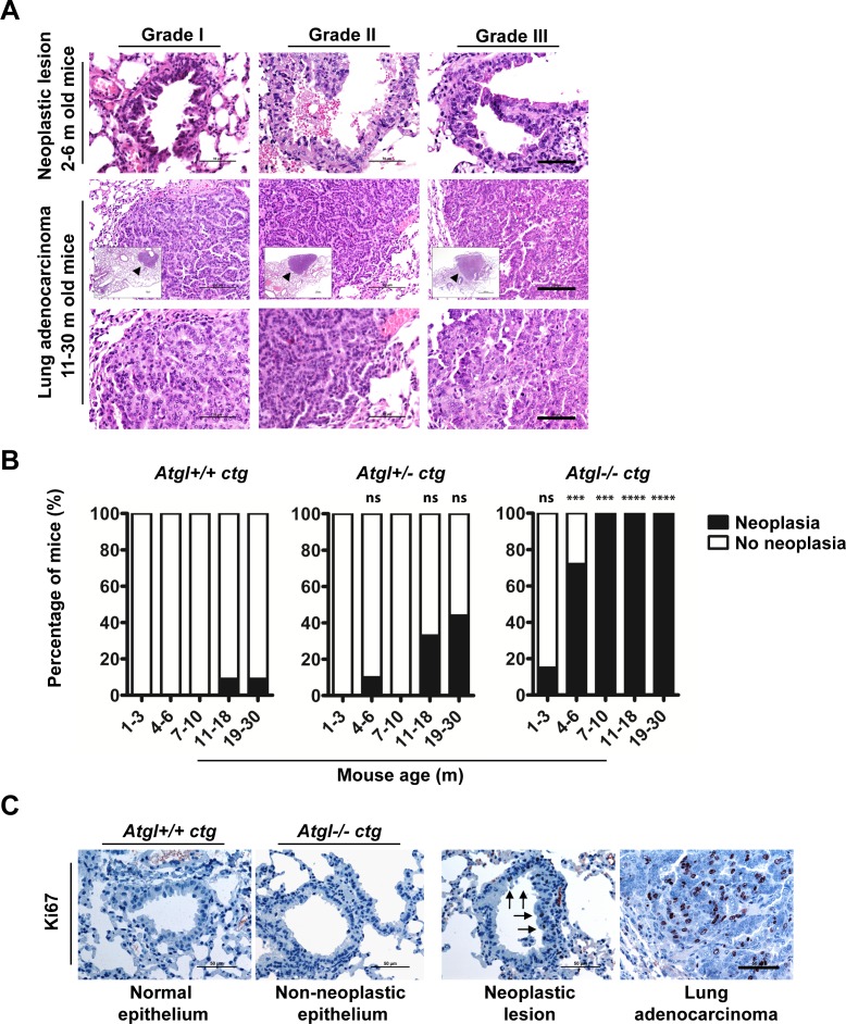
Metabolic reprogramming is a hallmark of cancer. Understanding cancer metabolism is instrumental to devise innovative therapeutic approaches. Anabolic metabolism, including the induction of lipogenic enzymes, is a key feature of proliferating cells. Here, we report a novel tumor suppressive function for adipose triglyceride lipase (ATGL), the rate limiting enzyme in the triglyceride hydrolysis cascade.In immunohistochemical analysis, non-small cell lung cancers, pancreatic adenocarcinoma as well as leiomyosarcoma showed significantly reduced levels of ATGL protein compared to corresponding normal tissues. The ATGL gene was frequently deleted in various forms of cancers. Low levels of ATGL mRNA correlated with significantly reduced survival in patients with ovarian, breast, gastric and non-small cell lung cancers. Remarkably, pulmonary neoplasia including invasive adenocarcinoma developed spontaneously in mice lacking ATGL pointing to an important role for this lipase in controlling tumor development.Loss of ATGL, as detected in several forms of human cancer, induces spontaneous development of pulmonary neoplasia in a mouse model. Our results, therefore, suggest a novel tumor suppressor function for ATGL and contribute to the understanding of cancer metabolism. We propose to evaluate loss of ATGL protein expression for the diagnosis of malignant tumors. Finally, modulation of the lipolytic pathway may represent a novel therapeutic approach in the treatment of human cancer.

Keywords: adipose triglyceride lipase; cancer metabolism; diagnostic marker; lipolysis.


Conflict of interest statement

The authors disclose no potential conflicts of interest.

Figures

Figure 1
Figure 1. ATGL protein is frequently reduced in human cancer
A. Representative images show haematoxylin and eosin (H&E) staining and immunohistochemical analysis (IHC) using antibodies against ATGL. Normal tissues (upper panels) are compared to cancers (lower panels). H-Score of ATGL protein levels (diagrams) determined by IHC takes into account the proportion (P, 0-100) of positive cells and the respective intensity score (I, 0-3) according to the formula [(I(0)xP(0))+(I(1)xP(1))+(I(2)xP(2))+(I(3)xP(3))]. LUE, normal lung epithelium; LUAD, lung adenocarcinoma; LUSC, lung squamous cell carcinoma; PAE, normal pancreatic epithelium; PanIN, pancreatic intraepithelial neoplasia; PAAD, pancreatic adenocarcinoma; UTSM, normal uterus smooth muscle; LM, benign tumor of smooth muscle (leiomyoma); LMS, malignant tumor of smooth muscle (leiomyosarcoma). ****P < .0001. Error bars represent SD. (LUE: mean = 13.47; n =13. LUAD: mean = 17.31; n = 70. LUSC: mean = 5.623; n = 34. PAE: mean = 263.4; n = 34. PAAD: mean = 22.02; n = 47. PanIN: mean = 23.48; n = 66. UTSM: mean = 296.5; n =26. LMS: mean = 42.53; n = 83. LM: mean = 291.0; n = 42). Scale bar, 50μm. B. Representative images show H&E staining and IHC using antibodies against ATGL of preinvasive neoplastic precursor lesions and invasive adenocarcinoma of the pancreas. PanIN-1, pancreatic intraepithelial neoplasia (low grade); PanIN-3, pancreatic intraepithelial neoplasia (high grade); PAAD, pancreatic adenocarcinoma. Arrows point to neoplastic lesions and invasive adenocarcinoma; N, normal epithelium. Normal epithelium stain positive for ATGL. C. Representative images show H&E and IHC using antibodies against ATGL. Normal smooth muscle and leiomyoma cells stain positive for ATGL whereas leiomyosarcoma cells are negative. Scale bar, 50μm.
Figure 2
Figure 2. Loss of ATGL results in spontaneous pulmonary neoplasia
A. Histopathological analysis of intraepithelial neoplastic lesions and invasive epithelial tumors (adenocarcinoma) in lungs of Atgl+/− ctg and Atgl−/− ctg mice. We did not detect lung adenocarcinoma in any of the mice from the three genotypes that were younger than 10 months. In mice older than 10 month, no lung adenocarcinoma were detected in Atgl+/+ ctg mice (n = 22). Out of 20 Atgl−/− ctg mice, we detected adenocarcinoma in 5 mice (P value = .0182). We also detected adenocarcinoma in 4 out of 21 Atgl+/− ctg mice (P value = .0485). (Upper panels) show intraepithelial neoplastic lesions. Scale bar, 50 μm. (Middle panels) show invasive adenocarcinoma. (Inserts) show the entire tumor area (arrowheads). Scale bar, 100 μm. Representative tumor regions are shown at higher magnification in the (lower panels) demonstrating cellular atypia. Scale bar, 50 μm. B. Bar graphs show the percentage of mice in which one or more pulmonary neoplastic lesions were detected. Atgl+/+ ctg mice, (n = 50); Atgl+/− ctg mice (n = 49); Atgl−/− ctg mice (n = 49). C. IHC analysis of Atgl+/+ ctg, Atgl+/− ctg, or Atgl−/− ctg lung sections using antibodies against Ki67 (proliferation marker). Representative images show no Ki67 positive cells in non-neoplastic lung epithelium of Atgl+/+ ctg and Atgl−/− ctg mice, whereas neoplastic lesion and adenocarcinoma show noticeable positive nuclei. Arrows highlight Ki67 positive nuclei and point to the part of a bronchiole with an intraepithelial neoplastic lesion. Scale bar, 50 μm.

References

    1. Hanahan D, Weinberg RA. Hallmarks of cancer: the next generation. Cell. 2011;144:646–674. - PubMed
    1. Ward PS, Thompson CB. Metabolic reprogramming: a cancer hallmark even warburg did not anticipate. Cancer cell. 2012;21:297–308. - PMC - PubMed
    1. Cantor JR, Sabatini DM. Cancer cell metabolism: one hallmark, many faces. Cancer discovery. 2012;2:881–898. - PMC - PubMed
    1. DeBerardinis RJ, Lum JJ, Hatzivassiliou G, Thompson CB. The biology of cancer: metabolic reprogramming fuels cell growth and proliferation. Cell metabolism. 2008;7:11–20. - PubMed
    1. Vander Heiden MG, Cantley LC, Thompson CB. Understanding the Warburg effect: the metabolic requirements of cell proliferation. Science. 2009;324:1029–1033. - PMC - PubMed

MeSH terms

LinkOut - more resources