. 2017 Jan 1;525(1):116-139.
doi: 10.1002/cne.24044. Epub 2016 Jun 19.

Pattern of distribution of serotonergic fibers to the amygdala and extended amygdala in the rat

Affiliations

Pattern of distribution of serotonergic fibers to the amygdala and extended amygdala in the rat

Stephanie B Linley et al. J Comp Neurol. .

Abstract

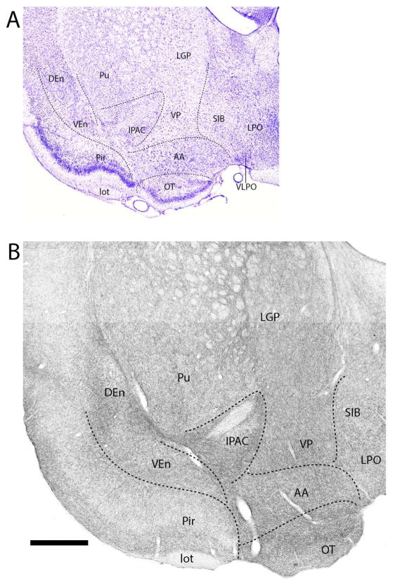
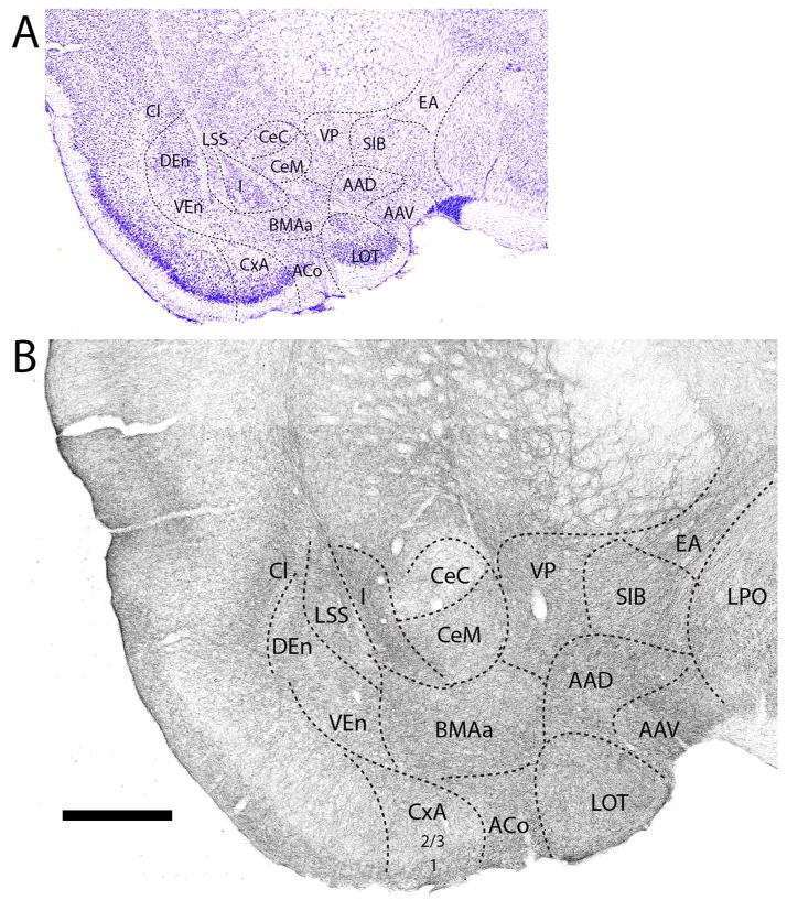
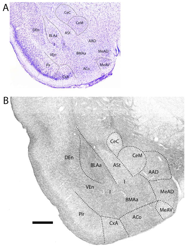
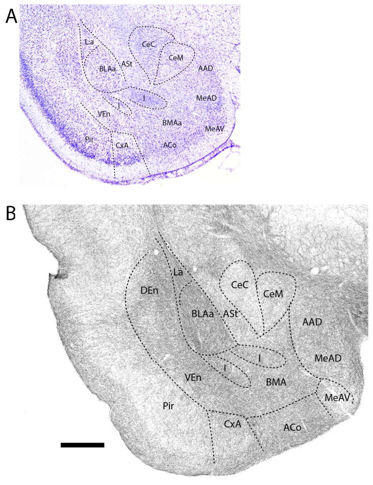
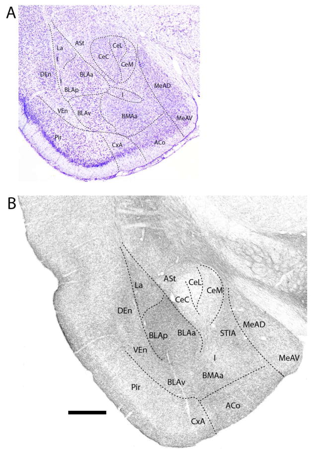
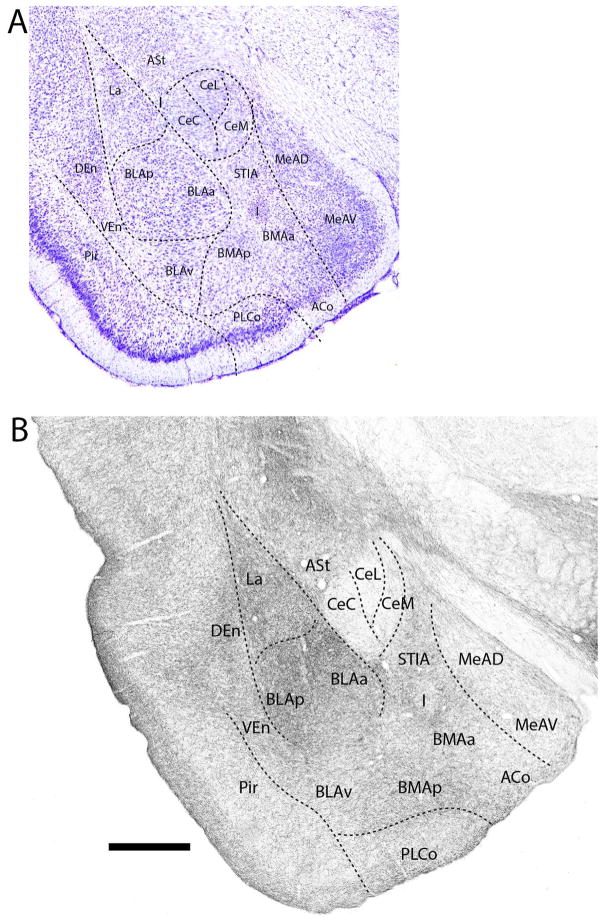
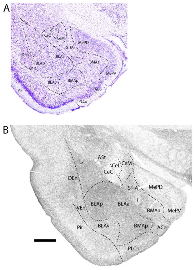
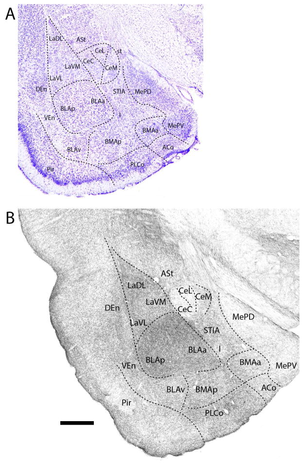
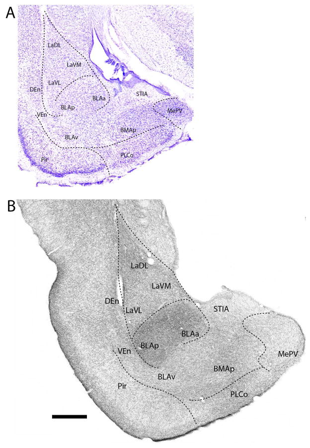
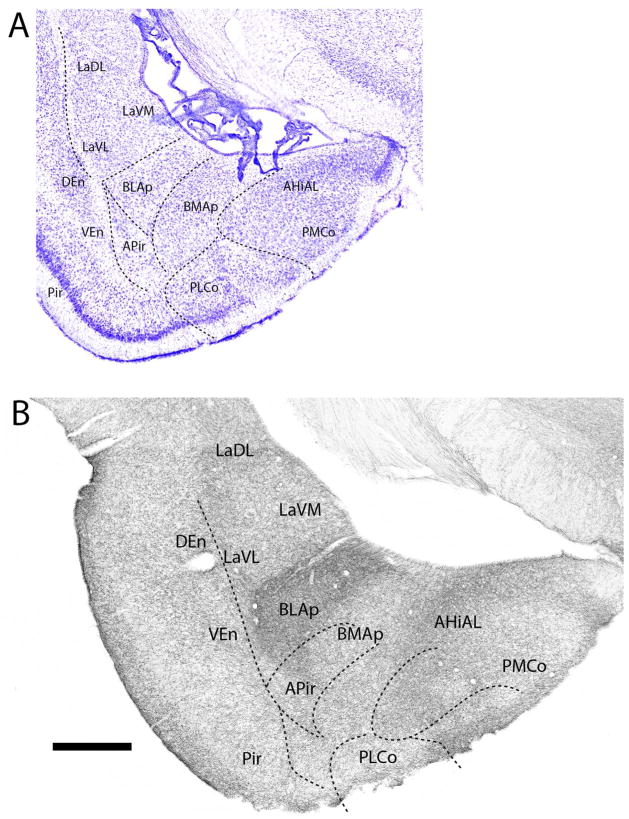
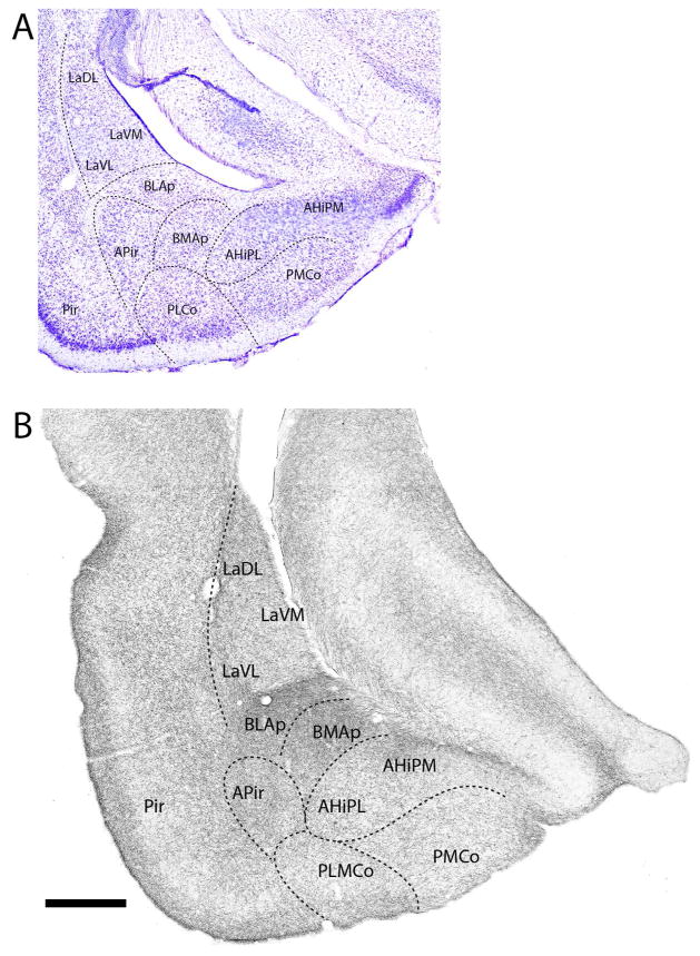
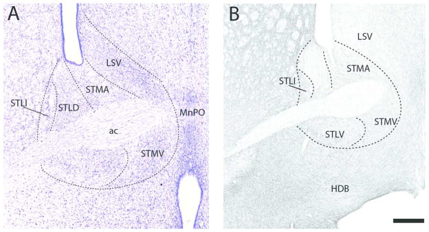
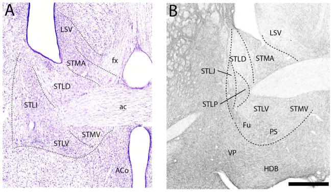
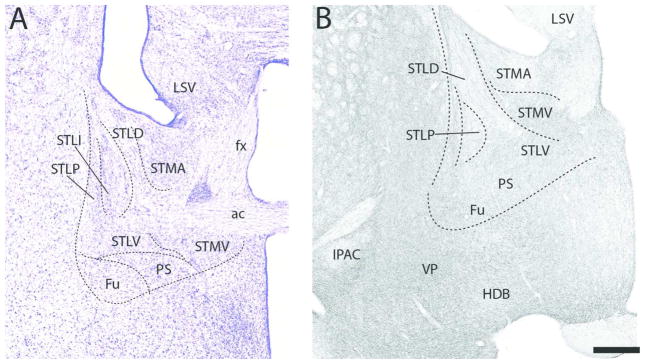
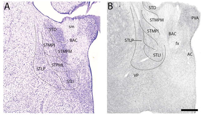
As is well recognized, serotonergic (5-HT) fibers distribute widely throughout the forebrain, including the amygdala. Although a few reports have examined the 5-HT innervation of select nuclei of the amygdala in the rat, no previous report has described overall 5-HT projections to the amygdala in the rat. Using immunostaining for the serotonin transporter, SERT, we describe the complete pattern of distribution of 5-HT fibers to the amygdala (proper) and to the extended amygdala in the rat. Based on its ontogenetic origins, the amygdala was subdivided into two major parts, pallial and subpallial components, with the pallial component further divided into superficial and deep nuclei (Olucha-Bordonau et al. 2015). SERT+ fibers were shown to distributed moderately to densely to the deep and cortical pallial nuclei, but, by contrast, lightly to the subpallial nuclei. Specifically, 1) of the deep pallial nuclei, the lateral, basolateral, and basomedial nuclei contained a very dense concentration of 5-HT fibers; 2) of the cortical pallial nuclei, the anterior cortical and amygdala-cortical transition zone rostrally and the posteromedial and posterolateral nuclei caudally contained a moderate concentration of 5-HT fibers; and 3) of the subpallial nuclei, the anterior nuclei and the rostral part of the medial (Me) nuclei contained a moderate concentration of 5-HT fibers, whereas caudal regions of Me as well as the central nuclei and the intercalated nuclei contained a sparse/light concentration of 5-HT fibers. With regard to the extended amygdala (primarily the bed nucleus of stria terminalis; BST), on the whole, the BST contained moderate numbers of 5-HT fibers, spread fairly uniformly throughout BST. The findings are discussed with respect to a critical serotonergic influence on the amygdala, particularly on the basal complex, and on the extended amygdala in the control of states of fear and anxiety. J. Comp. Neurol. 525:116-139, 2017. © 2016 Wiley Periodicals, Inc.

Keywords: 5-HT1A receptors; 5-HT2C receptors; anxiety; basolateral complex of amygdala; bed nucleus of stria terminalis; central nucleus of amygdala; fear; pallial amygdala; stress; subpallial amygdala.


Conflict of interest statement

STATEMENT The authors declare no conflict of interest.

Figures

Figure 1
Figure 1
A: Low magnification Nissl-stained transverse section at the transition between the amygdala (proper) and the extended amygdala showing the locations of nuclei at this level. B: Pattern of distribution of 5-HT fibers to nuclei of the amygdala at the same level – or to sublenticular region of the amygdala. See list for abbreviations. Scale bar for A = 1.2 mm; for B = 500 μm.
Figure 2
Figure 2
A: Low magnification Nissl-stained transverse section through the anterior amygdala showing the locations of nuclei at the same level. B: Pattern of the distribution of 5-HT fibers to nuclei of the amygdala at this level (B). See list for abbreviations. Scale bar for A = 1.3 mm; for B = 500 μm.
Figure 3
Figure 3
A: Low magnification Nissl-stained transverse section through the anterior amygdala showing the locations of nuclei at this level. B: Pattern of distribution of 5-HT fibers to nuclei of the amygdala at the same level. See list for abbreviations. Scale bar for A = 800 μm; for B = 500 μm.
Figure 4
Figure 4
A: Low magnification Nissl-stained transverse section through the anterior amygdala showing the locations of nuclei at this level. B: Pattern of distribution of 5-HT fibers to nuclei of the amygdala at the same level. Note the dense labeling of nuclei of the basolateral complex (La, BMA, BLA) and the light labeling of divisions of the central nucleus (Ce). See list for abbreviations. Scale bar for A = 1 mm; for B = 500 μm.
Figure 5
Figure 5
A: Low magnification Nissl-stained transverse section through the mid-portion of the amygdala showing the locations of nuclei at this level. B: Pattern of distribution of 5-HT fibers to nuclei of the amygdala at the same level. Note the dense labeling of nuclei of the basolateral complex (La, BMA, BLA), particularly BLA, and the sparse labeling of divisions of the central nucleus (Ce). See list for abbreviations. Scale bar for A = 1 mm; for B = 500 μm.
Figure 6
Figure 6
A: Low magnification Nissl-stained transverse section through the mid-portion of the amygdala showing the locations of nuclei at this level. B: Pattern of distribution of 5-HT fibers to nuclei of the amygdala at the same level. Note the dense labeling of nuclei of the basolateral complex (La, BMA, BLA), particularly BLA, and the sparse labeling of divisions of the central nucleus (Ce). See list for abbreviations. Scale bar for A = 1 mm; for B = 500 μm.
Figure 7
Figure 7
A: Low magnification Nissl-stained transverse section through the mid-portion of the amygdala showing the locations of nuclei at this level. B: Pattern of distribution of 5-HT fibers to nuclei of the amygdala at the same level. Note the dense labeling of nuclei of the basolateral complex (La, BMA, BLA), particularly BLA, and the sparse to light labeling of divisions of the central (Ce) and medial nucleus (Me). See list for abbreviations. Scale bar for A = 835 μm; for B = 500 μm.
Figure 8
Figure 8
A: Low magnification Nissl-stained transverse section through the posterior amygdala showing the locations of nuclei at this level. B: Pattern of distribution of 5-HT fibers to nuclei of the amygdala at the same level. Note the dense labeling of nuclei of the basolateral complex (La, BMA, BLA), particularly BLA, and the sparse to light labeling of divisions of the central (Ce) and medial nuclei (Me). See list for abbreviations. Scale bar for A = 700 μm; for B = 500 μm.
Figure 9
Figure 9
A: Low magnification Nissl-stained transverse section through the posterior amygdala showing the locations of nuclei at this level. B: Pattern of distribution of 5-HT fibers to nuclei of the amygdala at the same level. Note the dense labeling of nuclei of the basolateral complex (La, BMA, BLA), particularly BLA, and the light labeling of the medial nucleus. See list for abbreviations. Scale bar for A = 670 μm; for B = 500 μm.
Figure 10
Figure 10
A: Low magnification Nissl-stained transverse section through the posterior amygdala showing the locations of nuclei at this level. B: Pattern of distribution of 5-HT fibers to nuclei of the amygdala at the same level. Note the dense labeling of the posterior part of the basolateral nucleus, setting it apart from surrounding structures. See list for abbreviations. Scale bar for A = 1 mm; for B = 500 μm.
Figure 11
Figure 11
A: Low magnification Nissl-stained transverse section through the posterior amygdala showing the locations of nuclei at this level. B: Pattern of distribution of 5-HT fibers to nuclei of the amygdala at the same level. Note the dense labeling of the posterior part of the basomedial and basolateral nuclei. See list for abbreviations. Scale bar for A = 750 μm; for B = 500 μm.
Figure 12
Figure 12
A: Low magnification Nissl-stained transverse section through the anterior part of the extended amygdala showing the locations of nuclei at this level. B: Pattern of distribution of 5-HT fibers to nuclei of the extended amygdala at the same level. See list for abbreviations. Scale bar for A = 380 μm; for B = 250 μm.
Figure 13
Figure 13
A: Low magnification Nissl-stained transverse section through the anterior part of the extended amygdala showing the locations of nuclei at this level. B: Pattern of distribution of 5-HT fibers to nuclei of the extended amygdala at the same level. See list for abbreviations. Scale bar for A = 360 μm; for B = 250 μm.
Figure 14
Figure 14
A: Low magnification Nissl-stained transverse section through the posterior part of the extended amygdala showing the locations of nuclei at this level. B: Pattern of distribution of 5-HT fibers to nuclei of the extended amygdala at the same level. See list for abbreviations. Scale bar for A = 375 μm; for B = 250 μm.
Figure 15
Figure 15
A: Low magnification Nissl-stained transverse section through the posterior part of the extended amygdala showing the locations of nuclei at this level. B: Pattern of distribution of 5-HT fibers to nuclei of the extended amygdala at the same level. See list for abbreviations. Scale bar for A = 275 μm; B = 250 μm.

References

    1. Alheid GF. Extended amygdala and basal forebrain. Ann NY Acad Sci. 2003;985:185–205. - PubMed
    1. Amano T, Duvarci S, Popa D, Paré D. The fear circuit revisited: contributions of the basal amygdala nuclei to conditioned fear. J Neurosci. 2011;31:15481–15489. - PMC - PubMed
    1. Asan E, Yilmazer-Hanke DM, Eliava M, Hantsch M, Lesch KP, Schmitt A. The corticotropin-releasing factor (CRF)-system and monoaminergic afferents in the central amygdala: investigations in different mouse strains and comparison with the rat. Neuroscience. 2005;131:953–967. - PubMed
    1. Asan E, Steinke M, Lesch KP. Serotonergic innervation of the amygdala: targets, receptors, and implications for stress and anxiety. Histochem Cell Biol. 2013;139:785–813. - PubMed
    1. Bauer EP. Serotonin in fear conditioning processes. Behav Brain Res. 2015;277:68–77. - PubMed