

Stages of the Inflammatory Response in Pathology and Tissue Repair after Intracerebral Hemorrhage
- PMID: 27214704
- PMCID: PMC4956485
- DOI: 10.1055/s-0036-1582132
Stages of the Inflammatory Response in Pathology and Tissue Repair after Intracerebral Hemorrhage
Abstract
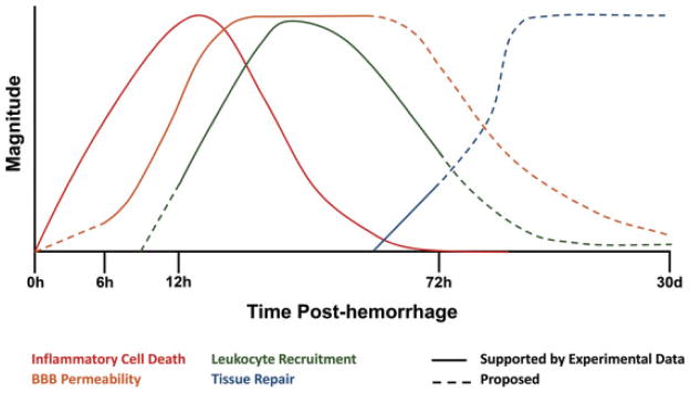
Intracerebral hemorrhage (ICH) is a major health concern, with high rates of mortality and morbidity and no highly effective clinical interventions. Basic research in animal models of ICH has provided insight into its complex pathology, in particular revealing the role of inflammation in driving neuronal death and neurologic deficits after hemorrhage. The response to ICH occurs in four distinct phases: (1) initial tissue damage and local activation of inflammatory factors, (2) inflammation-driven breakdown of the blood-brain barrier, (3) recruitment of circulating inflammatory cells and subsequent secondary immunopathology, and (4) engagement of tissue repair responses that promote tissue repair and restoration of neurologic function. The development of CNS inflammation occurs over many days after initial hemorrhage and thus may represent an ideal target for treatment of the disease, but further research is required to identify the mechanisms that promote engagement of inflammatory versus anti-inflammatory pathways. In this review, the authors examine how experimental models of ICH have uncovered critical mediators of pathology in each of the four stages of the inflammatory response, and focus on the role of the immune system in these processes.
Thieme Medical Publishers 333 Seventh Avenue, New York, NY 10001, USA.
Figures
References
-
- Mozaffarian D, Benjamin EJ, Go AS, et al. American Heart Association Statistics Committee and Stroke Statistics Subcommittee. Heart disease and stroke statistics—2015 update: a report from the American Heart Association. Circulation. 2015;131(4):e29–e322. - PubMed
-
- Aiyagari V. The clinical management of acute intracerebral hemorrhage. Expert Rev Neurother. 2015;15(12):1421–1432. - PubMed
-
- Felberg RA, Grotta JC, Shirzadi AL, et al. Cell death in experimental intracerebral hemorrhage: the “black hole” model of hemorrhagic damage. Ann Neurol. 2002;51(4):517–524. - PubMed
Publication types
MeSH terms
Substances
Grants and funding
LinkOut - more resources
Full Text Sources
Other Literature Sources
