. 2016 Jun 15;5(6):726-35.
doi: 10.1242/bio.018804.

SMN and coilin negatively regulate dyskerin association with telomerase RNA

Affiliations

SMN and coilin negatively regulate dyskerin association with telomerase RNA

Aaron R Poole et al. Biol Open. .

Abstract

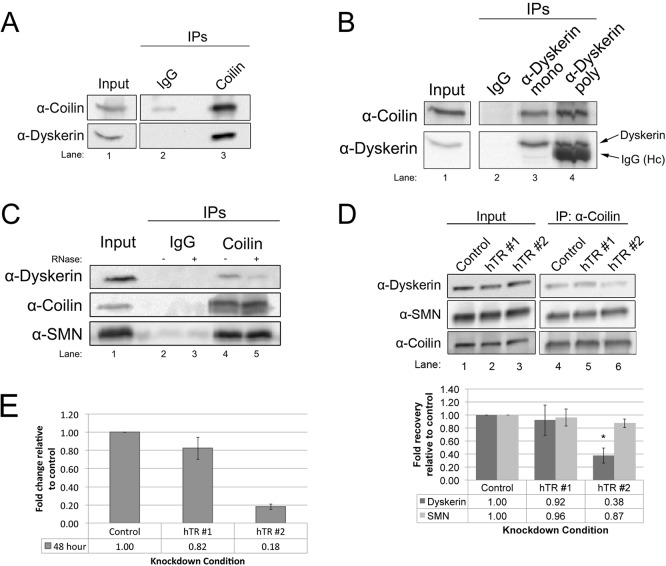
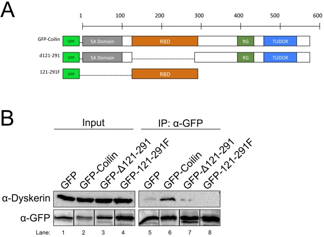
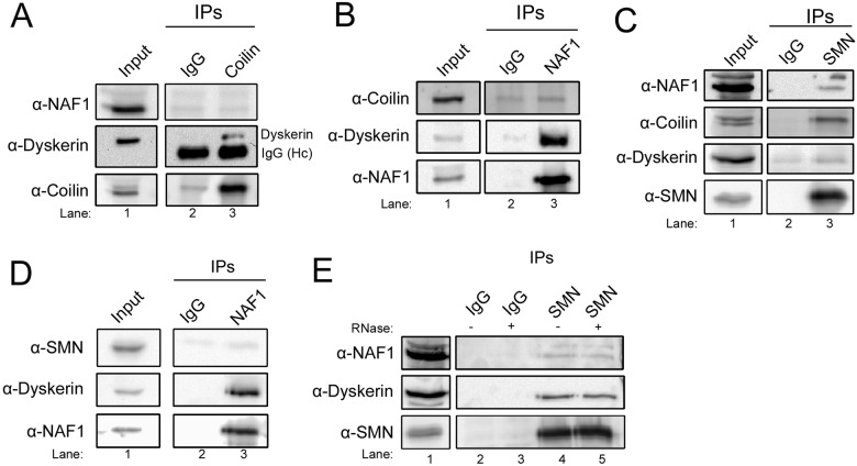
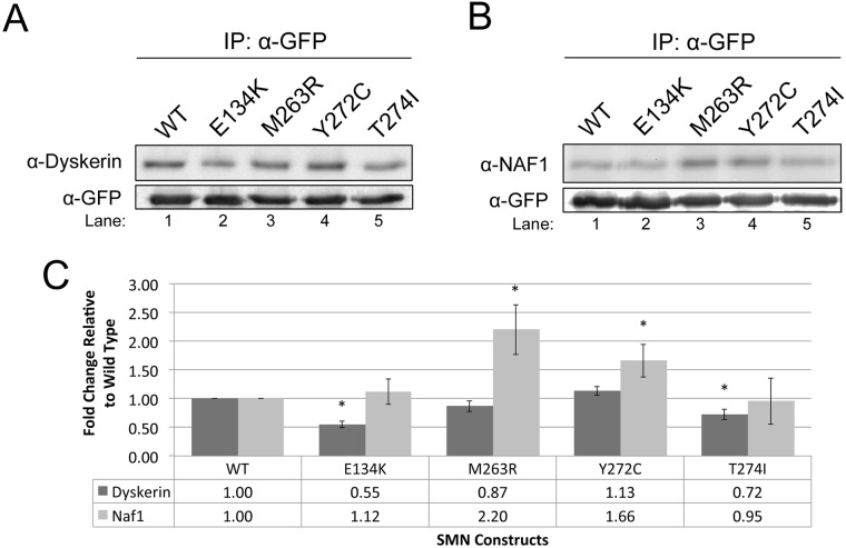
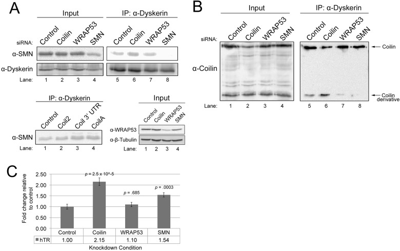
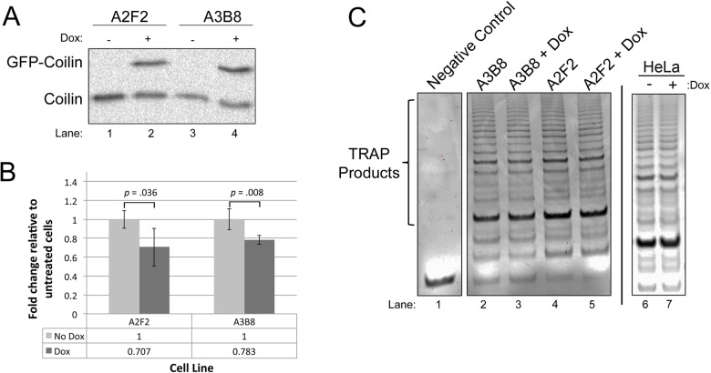
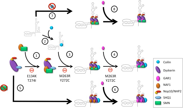
Telomerase is a ribonucleoprotein comprising telomerase RNA and associated proteins. The formation of the telomerase holoenzyme takes place in the Cajal body (CB), a subnuclear domain that participates in the formation of ribonucleoproteins. CBs also contribute to the delivery of telomerase to telomeres. The protein WRAP53 is enriched within the CB and is instrumental for the targeting of telomerase RNA to CBs. Two other CB proteins, SMN and coilin, are also suspected of taking part in some aspect of telomerase biogenesis. Here we demonstrate newly discovered associations between SMN and coilin with telomerase components, and further show that reduction of SMN or coilin is correlated with increased association of telomerase RNA with one these components, dyskerin. These findings argue that SMN and coilin may negatively regulate the formation of telomerase. Furthermore, clinically defined SMN mutants found in individuals with spinal muscular atrophy are altered in their association with telomerase complex proteins. Additionally, we observe that a coilin derivative also associates with dyskerin, and the amount of this protein in the complex is regulated by SMN, WRAP53 and coilin levels. Collectively, our findings bolster the link between SMN, coilin and the coilin derivative in the biogenesis of telomerase.

Keywords: Cajal body; Coilin; SMN; Telomerase.


Conflict of interest statement

Competing interests

The authors declare no competing or financial interests.

Figures

Fig. 1.
Fig. 1.
Coilin interaction with the telomerase complex protein dyskerin is mediated by hTR. (A) Lysate was immunoprecipitated (IPed) with either control (rabbit IgG) or coilin polyclonal antibodies, followed by running the samples on SDS-PAGE and western transfer. The probing antibodies are indicated. (B) Lysate was IPed with control (mouse IgG), dyskerin monoclonal or dyskerin polyclonal antibodies. Probing antibodies are indicated (Hc in lane 4=IgG heavy chain). (C) Lysate was IPed with control (rabbit IgG) or coilin polyclonal antibodies. Antibody-bead complexes were untreated (−) or treated (+) with RNase. Probing antibodies are indicated. (D) Cells were transfected with two different hTR siRNAs, followed by IP with anti-coilin antibodies. Probing antibodies are indicated. Quantification of dykserin and SMN recovery relative to that obtained with control siRNA for this and other experimental replicates is shown in the lower panel. For all IPs, inputs represent 1.5% of the cell lysate used in the IP reactions. (E) HeLa cells were treated with control or hTR siRNA (hTR #1 or hTR #2) for 48 h. Total RNA was collected and the amount of hTR was analyzed by qRT-PCR, relative to GAPDH message. The level of hTR in cells treated with hTR siRNA was compared to that found in cells with control siRNA for each time point (n=3). *P<0.05 compared to control knockdown. For all histograms, error bars denote standard error about the mean.
Fig. 2.
Fig. 2.
Mapping of the interaction domain on coilin for association with the dyskerin complex. (A) Schematic representation of coilin showing the locations of the self-association domain (SA Domain), RNA binding domain (RBD), RG-Box, and TUDOR-like domain (TUDOR) of the various GFP-tagged coilin constructs. (B) Cells were transfected with empty GFP vector or GFP-tagged coilin constructs, followed by IP with antibodies to GFP. Probing antibodies are indicated. For the GFP signals, the same membrane probed with anti-dyskerin was also probed with polyclonal anti-GFP antibodies. The bands for each GFP-tagged protein were then grouped together (lower panel). The experiment was repeated four times, and a representative result is shown. For all IPs, inputs represent 1.5% of cell lysate used in the IP reaction.
Fig. 3.
Fig. 3.
Differential interaction of coilin and SMN with telomerase complex proteins. (A) Lysate was IPed with either control (rabbit IgG) or coilin polyclonal antibodies. Probing antibodies are indicated. (B) IP of lysate with either control (rabbit IgG) or NAF1 polyclonal antibodies. Probing antibodies are indicated. (C) Lysate was IPed with either control (mouse IgG) or SMN monoclonal antibodies. Probing antibodies are indicated. (D) The reciprocal experiment of C was performed by IP of lysate with either control (rabbit IgG) or NAF1 polyclonal antibodies. Probing antibodies are indicated. (E) Lysate was IPed with either control (mouse IgG) or SMN monoclonal antibodies. The antibody-bead complexes were untreated (−) or treated (+) with RNase. Probing antibodies are indicated.
Fig. 4.
Fig. 4.
Clinically defined SMN mutants show alterations in the association with telomerase complex proteins. (A-B) Cells were transfected with wild-type (WT) or clinically defined SMN mutations, fused to GFP. IPs were performed using anti-GFP antibody. The resulting immunocomplexes were then probed for either dyskerin (A) or NAF1 (B). (C) Graphical representation of the data shown in (A) and (B) (n=8), normalized to the recovery of dyskerin and NAF1 by WT SMN. *P<0.05 compared to WT SMN, which serves as a control. Error bars represent standard error about the mean. For all IPs, inputs represent 1.5% the amount of the lysate used in the IP reaction. Note that GFP vector alone does not recover significant amounts of dyskerin (Fig. 2B, lane 5).
Fig. 5.
Fig. 5.
Reduction of Cajal body proteins alters the composition of the dyskerin complex. (A, upper panel) siRNA-treated lysate was IPed with anti-dyskerin. A portion of the IPs were subjected to SDS-PAGE, western blotting, and probing with anti-SMN or anti-dyskerin antibodies, while another portion of the IPs was used to isolate RNA for the experiment shown in C. (Lower panel) Three different siRNAs (Coil2, Coilin 3′ UTR or CoilA) were used to knockdown coilin in HeLa cells. Cells were also transfected with control siRNA. 48 h later, lysates were generated and IPed using a dyskerin monoclonal antibody. The IPs were then subjected to SDS-PAGE and western blotting. The membrane was then probed with a SMN antibody. Also shown is the knockdown of WRAP53 we achieve using WRAP53 siRNA compared to control, coilin or SMN siRNA. Western blot was probed with anti-WRAP53 then anti-tubulin antibodies. (B) siRNA-transfected cell lysate was IPed with anti-dyskerin antibody. The IPs were subjected to SDS-PAGE, western blotting and probing with anti-coilin antibodies. The location of full-length coilin and the 28 kDa coilin derivative is indicated. For all IPs, inputs represent 1.5% the amount of the lysate used in the IP reaction. (C) Coilin and SMN negatively regulate hTR association with dyskerin. siRNA-transfected lysate was IPed with dyskerin polyclonal antibody, followed by RNA isolation and qRT-PCR to determine the level of hTR, normalized to that obtained with control siRNA (P-values are shown, n=4 experimental sets). Error bars represent standard error about the mean.
Fig. 6.
Fig. 6.
Coilin overexpression is correlated with decreased association of hTR with dyskerin and reduced telomerase activity. (A) Verification of Dox induction of GFP-coilin in the A2F2 and A3B8 lines. Cells were untreated or treated for 48 h with Dox, followed by lysate generation, SDS-PAGE, western blotting and probing with anti-coilin antibodies. (B) Lysate from untreated or Dox-treated (48 h) A2F2 or A3B8 cells was IPed with anti-dyskerin, followed by RNA isolation and qRT-PCR to determine the relative amount of hTR. There is a significant decrease in hTR with dox induction compared to uninduced (P=0.035 for A2F2 line, P=0.008 for A3B8 line, n=4). Error bars represent standard error about the mean. (C) Telomerase activity assays were conducted on equal protein amounts from Dox-induced or uninduced lysate from A2F2 and A3B8 lines, and samples were run on an agarose gel and stained with ethidium bromide (lysis buffer served as negative control). Dox induction of GFP-coilin in A2F2 (lanes 4 and 5) resulted in a 20% decrease in the amount of TRAP signal compared to untreated (n=5, P<0.0005). For the A3B8 line (lanes 2 and 3), Dox induction resulted in a 10% decrease in TRAP signal compared to untreated (n=3, P<0.05). Note that the treatment of non-inducible HeLa cells with Dox did not alter telomerase activity compared to untreated cells (C, right panel).
Fig. 7.
Fig. 7.
A model for the negative regulation of SMN and coilin on telomerase biogenesis. (1) NAF1 and SMN form a complex which is then loaded on to a pre-assembled dyskerin complex containing dyskerin, SHQ1, NHP2, and Nop10. However, the SMN mutations E134K and T274I inhibit this pathway. (2) Coilin binds to nascent hTR near dyskerin's binding site. (3) The dyskerin complex is then loaded onto hTR; however, coilin is not lost in this process and remains in the complex. SMN mutations M263R and Y272C inhibit the association of the dyskerin complex with hTR. (4) GAR1 is exchanged for NAF1 in the dyskerin complex. SMN mutations M263R and Y272C may also inhibit biogenesis at this step by blocking the association of GAR1 with the dyskerin complex. (5) If SMN expression is reduced, the dyskerin complex loads more readily on to hTR (indicated by a larger arrow), and (6) assembly of telomerase proceeds in the absence of SMN. (7) Similarly, if coilin expression is reduced, the dyskerin complex containing SMN will more readily bind hTR (indicated by a larger arrow), (8) and assembly will continue in the absence of coilin. Note: the stoichiometry shown here in regards to SMN and coilin in the telomerase complex is not intended to be definitive. In fact, interactions between SMN and coilin with telomerase components are most likely transient and require sub-stoichiometric amounts.

Similar articles

Cited by

References

    1. Andrade L. E. C., Tan E. M. and Chan E. K. L. (1993). Immunocytochemical analysis of the coiled body in the cell cycle and during cell proliferation. Proc. Natl. Acad. Sci. USA 90, 1947-1951. 10.1073/pnas.90.5.1947 - DOI - PMC - PubMed
    1. Bachand F., Boisvert F.-M., Cote J., Richard S. and Autexier C. (2002). The product of the survival of motor neuron (SMN) gene is a human telomerase-associated protein. Mol. Biol. Cell 13, 3192-3202. 10.1091/mbc.E02-04-0216 - DOI - PMC - PubMed
    1. Bertrandy S., Burlet P., Clermont O., Huber C., Fondrat C., Thierry-Mieg D., Munnich A. and Lefebvre S. (1999). The RNA-binding properties of SMN: deletion analysis of the zebrafish orthologue defines domains conserved in evolution. Hum. Mol. Genet. 8, 775-782. 10.1093/hmg/8.5.775 - DOI - PubMed
    1. Broome H. J. and Hebert M. D. (2012). In vitro RNase and nucleic acid binding activities implicate coilin in U snRNA processing. PLoS ONE 7, e36300 10.1371/journal.pone.0036300 - DOI - PMC - PubMed
    1. Broome H. J. and Hebert M. D. (2013). Coilin displays differential affinity for specific RNAs in vivo and is linked to telomerase RNA biogenesis. J. Mol. Biol. 425, 713-724. 10.1016/j.jmb.2012.12.014 - DOI - PMC - PubMed

LinkOut - more resources