. 2016 Sep;12(3):521-35.
doi: 10.1007/s11302-016-9518-3. Epub 2016 May 23.

Long noncoding NONRATT021972 siRNA normalized abnormal sympathetic activity mediated by the upregulation of P2X7 receptor in superior cervical ganglia after myocardial ischemia

Affiliations

Long noncoding NONRATT021972 siRNA normalized abnormal sympathetic activity mediated by the upregulation of P2X7 receptor in superior cervical ganglia after myocardial ischemia

Guihua Tu et al. Purinergic Signal. 2016 Sep.

Erratum in

Abstract

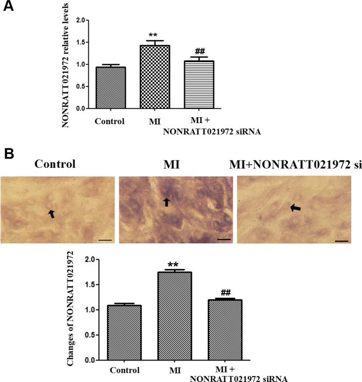
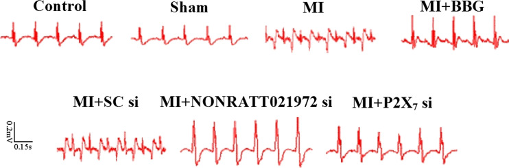
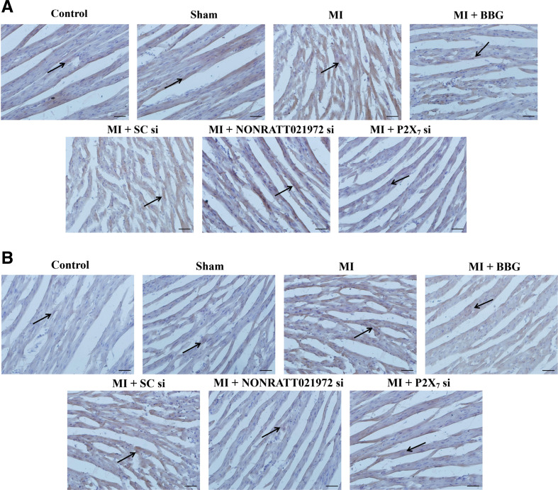
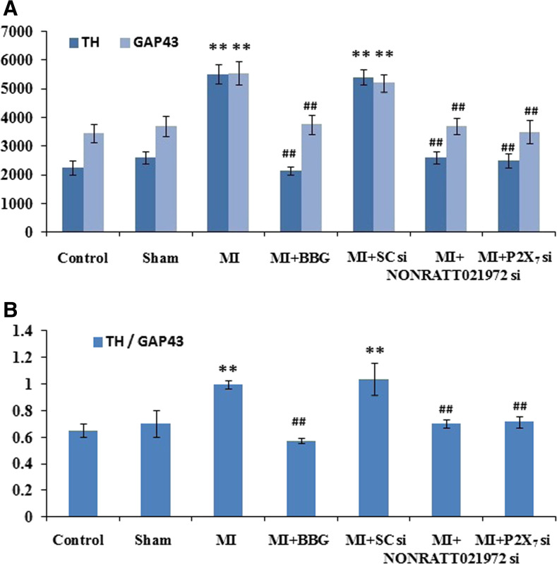
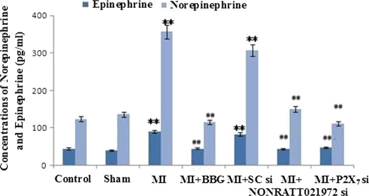
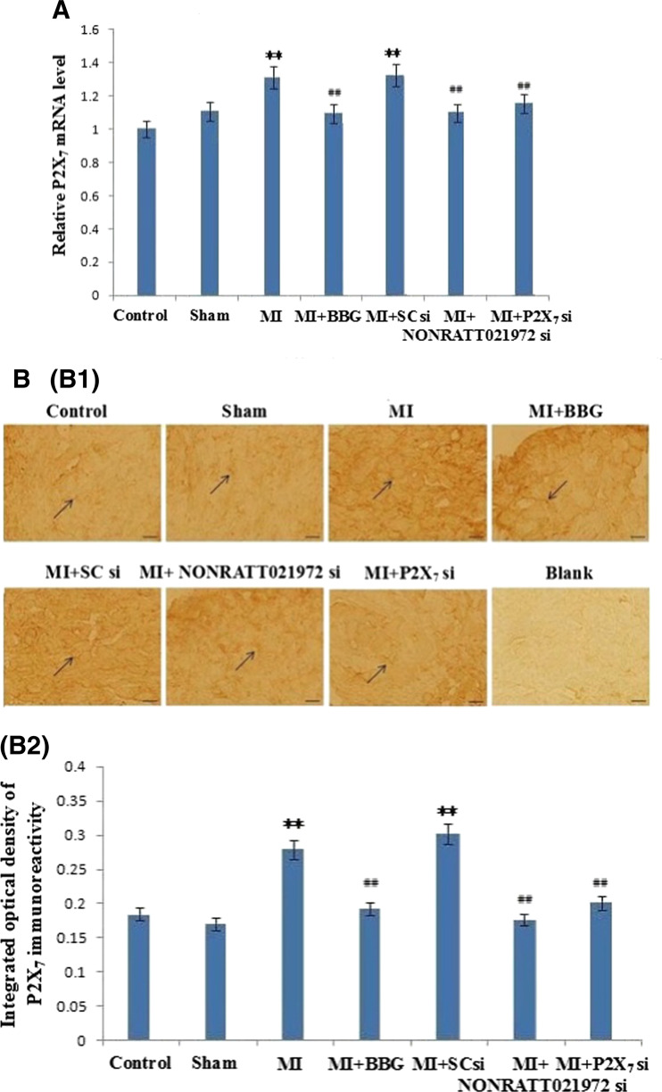
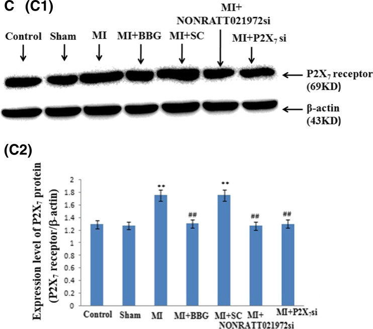
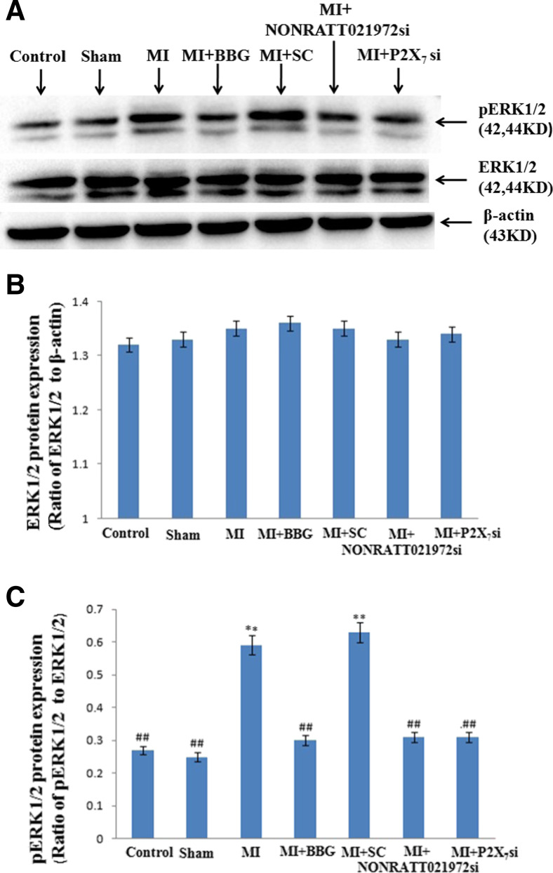
Previous studies showed that the upregulation of the P2X7 receptor in cervical sympathetic ganglia was involved in myocardial ischemic (MI) injury. The dysregulated expression of long noncoding RNAs (lncRNAs) participates in the onset and progression of many pathological conditions. The aim of this study was to investigate the effects of a small interfering RNA (siRNA) against the NONRATT021972 lncRNA on the abnormal changes of cardiac function mediated by the up-regulation of the P2X7 receptor in the superior cervical ganglia (SCG) after myocardial ischemia. When the MI rats were treated with NONRATT021972 siRNA, their increased systolic blood pressure (SBP), diastolic blood pressure (DBP), heart rate (HR), low-frequency (LF) power, and LF/HF ratio were reduced to normal levels. However, the decreased high-frequency (HF) power was increased. GAP43 and tyrosine hydroxylase (TH) are markers of nerve sprouting and sympathetic nerve fibers, respectively. We found that the TH/GAP43 value was significantly increased in the MI group. However, it was reduced after the MI rats were treated with NONRATT021972 siRNA. The serum norepinephrine (NE) and epinephrine (EPI) concentrations were decreased in the MI rats that were treated with NONRATT021972 siRNA. Meanwhile, the increased P2X7 mRNA and protein levels and the increased p-ERK1/2 expression in the SCG were also reduced. NONRATT021972 siRNA treatment inhibited the P2X7 agonist BzATP-activated currents in HEK293 cells transfected with pEGFP-P2X7. Our findings suggest that NONRATT021972 siRNA could decrease the upregulation of the P2X7 receptor and improve the abnormal changes in cardiac function after myocardial ischemia.

Keywords: Long noncoding RNA; Myocardial ischemia; P2X7 receptor; Superior cervical ganglia.


Conflict of interest statement

The authors declare that they have are no conflicts of interest.

Figures

Fig. 1
Fig. 1
The expression levels of NONRATT021972 in the SCG. a The expression levels of NONRATT021972 in the SCG were measured by quantitative real-time PCR, and the histogram shows the relative mRNA levels. The levels of NONRATT021972 expression in the SCG were significantly higher in the MI group than the con group. Treatment of the MI rats with NONRATT021972 siRNA decreased the expression of NONRATT021972 compared to MI group (P < 0.01). x¯±s, n = 3. **P < 0.01 compared to the con group. b The expression of NONRATT021972 in the SCG was significantly higher in the MI group than in the control group, as assessed by ISH (P < 0.01). Treatment of the MI rats with NONRATT021972 siRNA downregulated the expression of NONRATT021972 compared to MI group (P < 0.01). x¯±s, n = 5. **p < 0.01 compared with con group; ## p < 0.01 compared with MI. Scale bar = 50 μm; arrow shows the signal of NONRATT021972
Fig. 2
Fig. 2
The changes in the ECGs in each group of rats. The ST segment in the ECGs of the myocardial ischemia rats was high upward. At 30 days after the MI injury, there was an obvious abnormal Q wave in the MI and MI + SC si groups. The abnormal changes in the ECGs in response to the MI injury were greatly improved after the animals were treated with BBG, NONRATT021972 siRNA, or P2X7 siRNA
Fig. 3
Fig. 3
The density of cardiac sympathetic nerve fibers in each group of rats. The densities of GAP43-positive (a, c) and TH-positive (b, c) fibers around the ischemic myocardium in the MI group were significantly higher than those in the con, sham, MI + BBG, MI + NONRATT021972 si, and MI + P2X7 si groups (P < 0.01), and the TH/GAP43 value (d) in the MI rats was significantly increased compared to those in the con, sham, MI + BBG, MI + NONRATT021972 si, and MI + P2X7 si groups (P < 0.01). The densities (integrated optical density (IOD)) of the GAP43- and TH-positive fibers around the ischemic myocardium of the MI group were significantly increased compared with the control group (P < 0.01). The TH/GAP43 value in the MI rats treated with NONRATT021972 siRNA group was significantly decreased compared with rats in the MI group (P < 0.01). This indicates that the regenerative nerve fibers were primarily sympathetic nerves. x¯±s, n = 6. **P < 0.01, compared to the con group; ## P < 0. 01 vs the MI group. Scale bars 50 μm
Fig. 3
Fig. 3
The density of cardiac sympathetic nerve fibers in each group of rats. The densities of GAP43-positive (a, c) and TH-positive (b, c) fibers around the ischemic myocardium in the MI group were significantly higher than those in the con, sham, MI + BBG, MI + NONRATT021972 si, and MI + P2X7 si groups (P < 0.01), and the TH/GAP43 value (d) in the MI rats was significantly increased compared to those in the con, sham, MI + BBG, MI + NONRATT021972 si, and MI + P2X7 si groups (P < 0.01). The densities (integrated optical density (IOD)) of the GAP43- and TH-positive fibers around the ischemic myocardium of the MI group were significantly increased compared with the control group (P < 0.01). The TH/GAP43 value in the MI rats treated with NONRATT021972 siRNA group was significantly decreased compared with rats in the MI group (P < 0.01). This indicates that the regenerative nerve fibers were primarily sympathetic nerves. x¯±s, n = 6. **P < 0.01, compared to the con group; ## P < 0. 01 vs the MI group. Scale bars 50 μm
Fig. 4
Fig. 4
The concentrations of NE and EPI in the rats’ sera were measured by ELISA. The serum concentrations of NE and EPI in the MI group were significantly higher than those in the con, sham, MI + BBG, MI + NONRATT021972 si, and MI + P2X7 si groups (P < 0.01). The serum concentrations of NE and EPI in the MI group were significantly increased compared with the control group (P < 0.01). The serum concentrations of NE and EPI in the MI rats treated with NONRATT021972 siRNA group were significantly decreased compared with rats in the MI group (P < 0.01). There were no differences between the con, sham, MI + BBG, MI + NONRATT021972 si, and MI + P2X7 si groups (P > 0.05), or between the MI and MI + SC si groups (P > 0.05). x¯±s, n = 6. **P < 0.01, compared to the con group; ## P < 0. 01 vs the MI group
Fig. 5
Fig. 5
The levels of the P2X7 receptor mRNA, immunoreactivity, and protein in the SCG. a The histogram showed the relative mRNA levels were measured by quantitative real-time PCR. The levels of the P2X7 receptor mRNA in the MI and MI + SC si groups were significantly higher than those in the con, sham, MI + BBG, MI + NONRATT021972 si, and MI + P2X7 si groups (P < 0.01). The intensity of the P2X7 mRNA transcripts in the MI group was significantly increased compared with the control group (P < 0.01). The levels of the P2X7 mRNA transcripts in the MI rats treated with NONRATT021972 siRNA group were significantly decreased compared with rats in the MI group (P < 0.01). There were no differences between the con, sham, MI + BBG, MI + NONRATT021972 si, and MI + P2X7 si group (P > 0.05), or between the MI and MI + SC si groups (P > 0.05). BBG, NONRATT021972 siRNA, and P2X7 siRNA reduced the levels of the P2X7 receptor mRNA in the myocardial ischemia rats. x¯±s, n = 3. **P < 0.01 compared to the Con group; ## P < 0. 01 vs the MI group. b P2X7 immunoreactivity in the SCG of each of group rats was tested by immunohistochemistry: representative photos of P2X7 immunoreactivity in the SCG (B1); the histogram showed the results of the integrated optical density (IOD) for each group (B2). The intensity of P2X7 immunoreactivity in the MI group was obviously increased compared to the con, sham, MI + BBG, MI + NONRATT021972 si, and MI + P2X7 si groups (P < 0.01). The intensity of the P2X7 immunoreactivity in the MI group was significantly increased compared with the control group (P < 0.01). The intensity of the P2X7 immunoreactivity in the MI rats treated with NONRATT021972 siRNA group were significantly decreased compared with rats in the MI group (P < 0.01). There were no differences between the con, sham, MI + BBG, MI + NONRATT021972 si, and MI + P2X7 si groups (P > 0.05), or between the MI and MI + SC si groups (P > 0.05). The arrows indicate the immunostained neurons. x¯±s, n = 10. **P < 0.01 compared to the con group; ## P < 0. 01 vs the MI group. Scale bars 20 μm. c The expression of the P2X7 protein in the SCG of each group of rats was tested by Western blotting: representative blots of the P2X7 protein in the SCG (C1); the histogram showed the IOD of the results (C2). The IOD of P2X7 receptor expression in the MI group was obviously increased compared to the con, sham, MI + BBG, MI + NONRATT021972 si, and MI + P2X7 si groups (P < 0.01). The IOD of the P2X7 protein expression in the MI group was significantly increased compared with the control group (P < 0.01). The IOD of the P2X7 protein in the MI rats treated with NONRATT021972 siRNA group were significantly decreased compared with rats in the MI group (P < 0.01). There were no differences between the con, sham, MI + BBG, MI + NONRATT021972 si, and MI + P2X7 si groups (P > 0.05), or between the MI and MI + SC si groups (P > 0.05). x¯±s, n = 6. **P < 0.01, compared to the con group; ## P < 0.01 vs the MI group
Fig. 5
Fig. 5
The levels of the P2X7 receptor mRNA, immunoreactivity, and protein in the SCG. a The histogram showed the relative mRNA levels were measured by quantitative real-time PCR. The levels of the P2X7 receptor mRNA in the MI and MI + SC si groups were significantly higher than those in the con, sham, MI + BBG, MI + NONRATT021972 si, and MI + P2X7 si groups (P < 0.01). The intensity of the P2X7 mRNA transcripts in the MI group was significantly increased compared with the control group (P < 0.01). The levels of the P2X7 mRNA transcripts in the MI rats treated with NONRATT021972 siRNA group were significantly decreased compared with rats in the MI group (P < 0.01). There were no differences between the con, sham, MI + BBG, MI + NONRATT021972 si, and MI + P2X7 si group (P > 0.05), or between the MI and MI + SC si groups (P > 0.05). BBG, NONRATT021972 siRNA, and P2X7 siRNA reduced the levels of the P2X7 receptor mRNA in the myocardial ischemia rats. x¯±s, n = 3. **P < 0.01 compared to the Con group; ## P < 0. 01 vs the MI group. b P2X7 immunoreactivity in the SCG of each of group rats was tested by immunohistochemistry: representative photos of P2X7 immunoreactivity in the SCG (B1); the histogram showed the results of the integrated optical density (IOD) for each group (B2). The intensity of P2X7 immunoreactivity in the MI group was obviously increased compared to the con, sham, MI + BBG, MI + NONRATT021972 si, and MI + P2X7 si groups (P < 0.01). The intensity of the P2X7 immunoreactivity in the MI group was significantly increased compared with the control group (P < 0.01). The intensity of the P2X7 immunoreactivity in the MI rats treated with NONRATT021972 siRNA group were significantly decreased compared with rats in the MI group (P < 0.01). There were no differences between the con, sham, MI + BBG, MI + NONRATT021972 si, and MI + P2X7 si groups (P > 0.05), or between the MI and MI + SC si groups (P > 0.05). The arrows indicate the immunostained neurons. x¯±s, n = 10. **P < 0.01 compared to the con group; ## P < 0. 01 vs the MI group. Scale bars 20 μm. c The expression of the P2X7 protein in the SCG of each group of rats was tested by Western blotting: representative blots of the P2X7 protein in the SCG (C1); the histogram showed the IOD of the results (C2). The IOD of P2X7 receptor expression in the MI group was obviously increased compared to the con, sham, MI + BBG, MI + NONRATT021972 si, and MI + P2X7 si groups (P < 0.01). The IOD of the P2X7 protein expression in the MI group was significantly increased compared with the control group (P < 0.01). The IOD of the P2X7 protein in the MI rats treated with NONRATT021972 siRNA group were significantly decreased compared with rats in the MI group (P < 0.01). There were no differences between the con, sham, MI + BBG, MI + NONRATT021972 si, and MI + P2X7 si groups (P > 0.05), or between the MI and MI + SC si groups (P > 0.05). x¯±s, n = 6. **P < 0.01, compared to the con group; ## P < 0.01 vs the MI group
Fig. 6
Fig. 6
The expression of ERK1/2 and p-ERK 1/2 receptor in the SCG of each group was tested by Western blotting. a Representative photos of the levels of the ERK1/2 and p-ERK1/2 proteins in the SCG. b The histogram showed the ratio of ERK1/2 to β-actin. c The histogram showed the ratio of p ERK1/2 to ERK1/2. The integrated optical density (IOD) of p-ERK1/2 expression in the MI group was obviously increased compared to the con, sham, MI + BBG, MI + NONRATT021972 si, and MI + P2X7 si groups (P < 0.01). The IOD of p-ERK1/2 expression in the MI group was significantly increased compared with the control group (P < 0.01). The IOD of p-ERK1/2 expression in the MI rats treated with NONRATT021972 siRNA group was significantly decreased compared with rats in the MI group (P < 0.01). There were no differences between the con, sham, MI + BBG, MI + NONRATT021972 si, and MI + P2X7 si groups (P > 0.05), or between the MI and MI + SC si groups (P > 0.05). x¯±s , n = 6. ** P < 0.01, compared to the con group; ## P < 0.01 vs the MI group
Fig. 7
Fig. 7
Effects of the NONRATT021972 siRNA on the P2X7 receptor agonist-activated current in HEK293 cells. Whole-cell patch-clamp recordings were performed on HEK293 cells expressing hP2X7. Inward currents were evoked by 100 μM BzATP at a holding potential of −60 mV. a BzATP-activated currents were recorded in HEK293 cells transfected with hP2X7 alone or cotransfected with the NONRATT021972 siRNA. b The histogram shows the normalized, mean peak current (mean ± S, n = 12, **P < 0.01, compared to the con group; ## P < 0.01 vs the MI group). NONRATT021972 siRNA decreased the BzATP-induced currents in HEK293 cells expressing the hP2X7 receptor

References

    1. Ponting CP, Belgard TG. Transcribed dark matter: meaning or myth? Hum Mol Genet. 2010;19(R2):R162–R168. doi: 10.1093/hmg/ddq362. - DOI - PMC - PubMed
    1. Kapranov P, Cheng J, Dike S, Nix DA, Duttagupta R, Willingham AT, Stadler PF, Hertel J, Hackermuller J, Hofacker IL, Bell I, Cheung E, Drenkow J, Dumais E, Patel S, Helt G, Ganesh M, Ghosh S, Piccolboni A, Sementchenko V, Tammana H, Gingeras TR. RNA maps reveal new RNA classes and a possible function for pervasive transcription. Science. 2007;316(5830):1484–1488. doi: 10.1126/science.1138341. - DOI - PubMed
    1. Huang Y, Liu N, Wang JP, Wang YQ, Yu XL, Wang ZB, Cheng XC, Zou Q. Regulatory long non-coding RNA and its functions. J Physiol Biochem. 2012;68(4):611–618. doi: 10.1007/s13105-012-0166-y. - DOI - PMC - PubMed
    1. Vance KW, Ponting CP. Transcriptional regulatory functions of nuclear long noncoding RNAs. Trends Genet. 2014;30(8):348–355. doi: 10.1016/j.tig.2014.06.001. - DOI - PMC - PubMed
    1. Rinn JL, Chang HY. Genome regulation by long noncoding RNAs. Annu Rev Biochem. 2012;81:145–166. doi: 10.1146/annurev-biochem-051410-092902. - DOI - PMC - PubMed

Publication types

MeSH terms

Substances

LinkOut - more resources