Review
. 2016 May 27:67:691-717.
doi: 10.1146/annurev-physchem-031215-010949.

Fundamental Properties of One-Dimensional Zinc Oxide Nanomaterials and Implementations in Various Detection Modes of Enhanced Biosensing

Affiliations
Review

Fundamental Properties of One-Dimensional Zinc Oxide Nanomaterials and Implementations in Various Detection Modes of Enhanced Biosensing

Jong-In Hahm. Annu Rev Phys Chem. .

Abstract

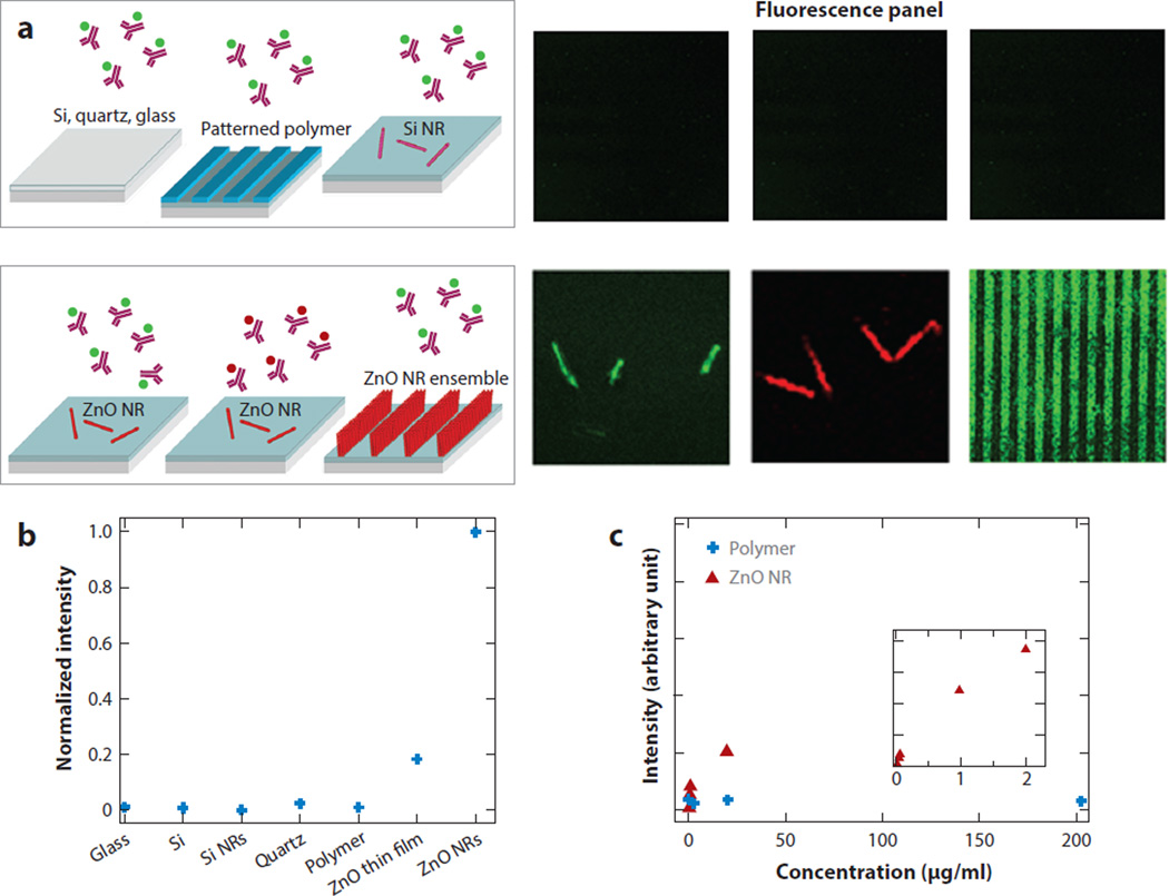
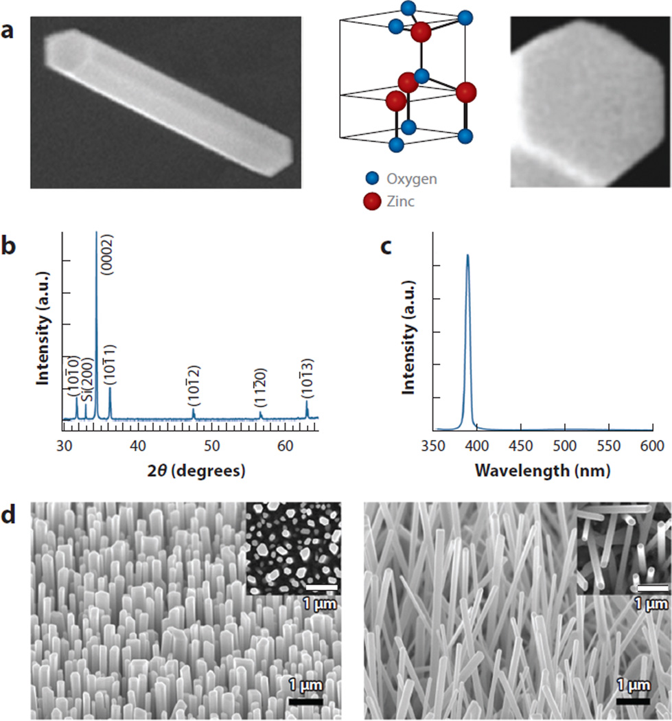
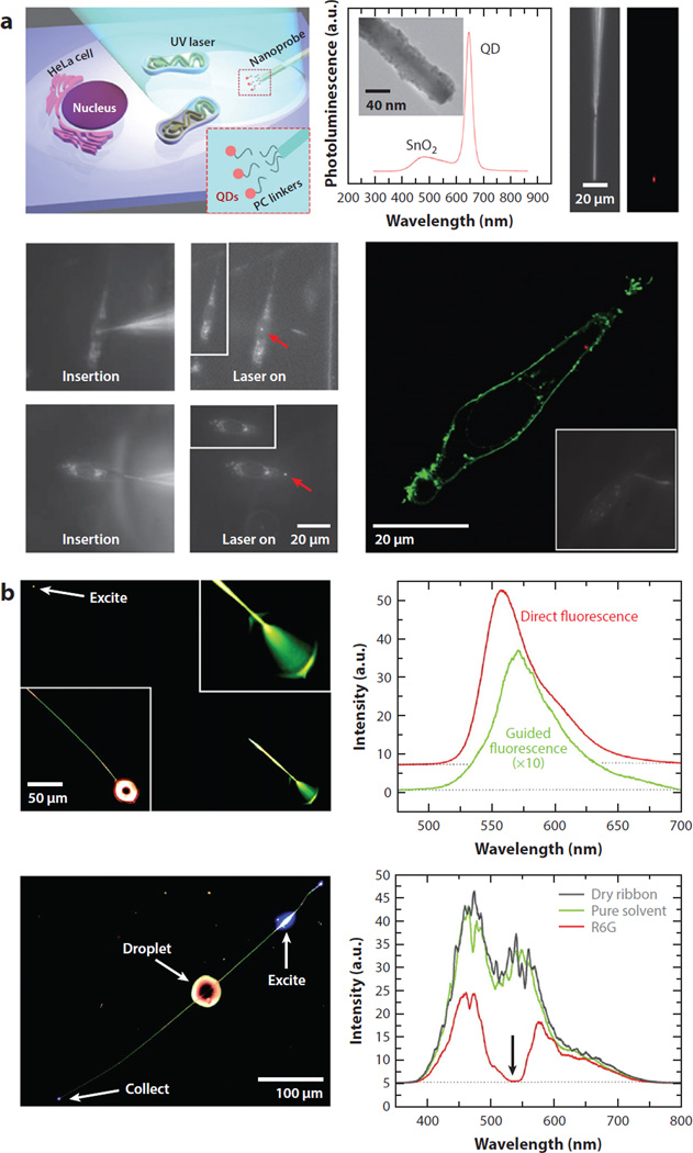
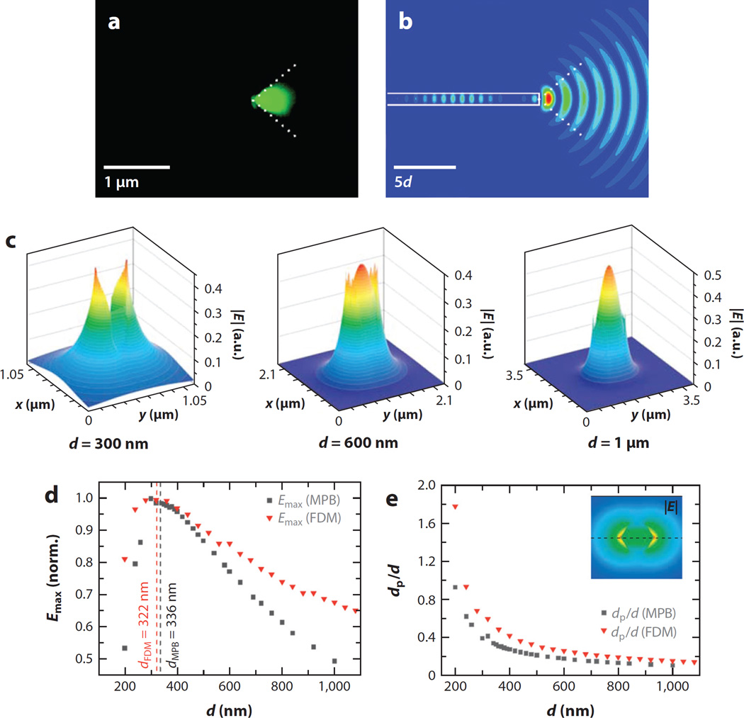
Recent bioapplications of one-dimensional (1D) zinc oxide (ZnO) nanomaterials, despite the short development period, have shown promising signs as new sensors and assay platforms offering exquisite biomolecular sensitivity and selectivity. The incorporation of 1D ZnO nanomaterials has proven beneficial to various modes of biodetection owing to their inherent properties. The more widely explored electrochemical and electrical approaches tend to capitalize on the reduced physical dimensionality, yielding a high surface-to-volume ratio, as well as on the electrical properties of ZnO. The newer development of the use of 1D ZnO nanomaterials in fluorescence-based biodetection exploits the innate optical property of their high anisotropy. This review considers stimulating research advances made to identify and understand fundamental properties of 1D ZnO nanomaterials, and examines various biosensing modes utilizing them, while focusing on the unique optical properties of individual and ensembles of 1D ZnO nanomaterials specifically pertaining to their bio-optical applications in simple and complex fluorescence assays.

Keywords: biomedical detection; biosensing; enhanced fluorescence detection; nanorod optical property; optical signal enhancement; zinc oxide nanomaterial.


Figures

Figure 1
Figure 1
Comparison of fluorescence emission monitored from 200-µg/ml fluorescein isothiocyanate (FITC)-conjugated or tetramethyl rhodamine isothiocyanate (TRITC)-conjugated antibovine immunoglobulin G on ZnO nanorods (NRs) versus control substrates after identical biodeposition. (a) No discernable fluorescence signal is detected from the biomolecules on the control substrates, including glass, quartz, native SiO2/Si, Si NRs, and polymeric surfaces of polystyrene (PS) and polymethylmethacrylate (PMMA). Conversely, a strong fluorescence signal is observed from individual and striped ZnO NR platforms. (b) Normalized fluorescence intensities observed from the biomolecules on various substrates are compared. (c) The fluorescence intensity was measured as a function of protein concentration on PMMA (blue) and ZnO NRs (red). The background fluorescence from PS and PMMA platforms inherent to these materials was subtracted from the fluorescence intensity measured after biodeposition. Figure adapted from Reference with permission. Copyright 2006 American Chemical Society.
Figure 2
Figure 2
Gas-phase synthesized ZnO nanorods (NRs). (a) 763 nm × 1.2 µm and 200 nm × 200 nm scanning electron microscopy (SEM) images showing the side (prismic) and end (basal) facets of a wurtzite ZnO NR. The width and length of the ZnO NR are 180 nm and 1.2 µm, respectively. Panel a adapted from Reference with permission. Copyright 2008 American Chemical Society. (b) X-ray diffraction data displaying the high crystalline quality of as-synthesized ZnO NRs showing the pronounced peak at 2θ = 34.5° corresponding to the preferential growth direction along 〈0001〉. (c) Room-temperature photoluminescence spectrum showing the extremely strong and narrow band-edge emission of ZnO NRs at 390 nm with no significant defect emission in the visible range. Panels b and c adapted from Reference with permission. Copyright 2005 American Scientific Publishers. (d) SEM images of gas-phase-synthesized ZnO NRs revealing densely grown, vertically oriented ZnO NRs on the Si (100) growth substrate. (Insets) Plan-view images. Panel d adapted from Reference with permission. Copyright 2005 American Chemical Society.
Figure 3
Figure 3
Exemplar applications of 1D NRs in guiding and collecting light. (a) Spatiotemporal delivery of QDs into a living cell is attempted using a photoactivatable SnO2 NR endoscope. Fluorescence confocal image of a HeLa cell shows the QDs (red dots in the cytoplasm) within the cell membrane (green) successfully delivered by the nanoprobe. Panel a adapted from Reference with permission. Copyright 2012 Nature Publishing Group. (b) Fluorescence and absorbance spectra of R6G are recorded using an SnO2 NR waveguide (240 nm × 260 nm × 540 µm) excited at one end (with top panels showing fluorescence geometry) and near the middle of the NR (with bottom panels showing absorption geometry). Panel b adapted from Reference with permission. Copyright 2005 National Academy of Sciences, USA. Abbreviations: NR, nanorod; PC, photocleavable; QD, quantum dot; R6G, rhodamine 6G; UV, ultraviolet.
Figure 4
Figure 4
Evidence of guided mode emission through a ZnO nanorod (NR) and computer-simulated results of an increased evanescent wave field and its penetration depth for ZnO NRs of various diameters. (a) Conical 90° emission of a low-order guided mode observed from one end of a ZnO NR. (b) 2D finite-difference time-domain calculation of the square of the electric field of a light pulse emitted from a NR of diameter d. Panels a and b adapted from Reference with permission. Copyright 2007 American Chemical Society. (c) The electric field |E| of a ZnO NR graphed for NR diameters of 300 nm, 600 nm, and 1 µm at λ = 761 nm. (d) Normalized maximum magnitude of the evanescent field (Emax) plotted as a function of the NR diameter. (e) Penetration depth (dp) obtained by the MIT Photonic-Bands (MPB) program and the finite-difference method (FDM) as a function of diameter for λ = 761 nm. Panels c, d, and e adapted from Reference with permission. Copyright 2007 Wiley-VCH Verlag GmbH & Co. KGaA, Weinheim.
Figure 5
Figure 5
Highly spatially localized, temporally extended biomolecular fluorescence signal observed on single ZnO nanorods (NRs). The contour map and the time-lapse images display variations in the biomolecular fluorescence intensity along the long axis of a 25-µm-long ZnO NR. Differences in the time dependence of the fluorescence intensity decay under constant irradiation were clearly observed depending on the ZnO NR planes. Red and black data show biomolecular fluorescence measured from the NR end and main body, respectively. Figure adapted from Reference with permission. Copyright 2014 Royal Society of Chemistry.
Figure 6
Figure 6
Factors governing the occurrence, degree, and magnitude of fluorescence intensification on nanorod ends (FINE). (a) Finite-difference time-domain simulations were carried out to obtain the radiation patterns from a single emitter radiating at 576 nm polarized along the x (top), y (middle), and z (bottom) directions. (b) The dimensional effect of ZnO nanorods (NRs) on FINE was evaluated by simulating far-field radiation patterns of a 517-nm electric dipole. A pair of far-field patterns is shown for each NR of the specified length (L) and width (d), where the top/bottom simulation corresponds to the spatial patterns observed from the z/x axis. (c) ZnO NRs of various lengths and widths were analyzed after treatment with three different concentrations of 5-(4,6-dichlorotriazinyl) aminofluorescein (DTAF)–antibovine IgG (anti-IgG). In all cases, the normalized ΔI value of (Iavg,NRefIavg,NRsf) indicated that the degree of FINE increased as the NR length increased. (d) When the spatial and temporal emission behavior of 1-µg/ml TRITC-anti-IgG on a lateral (L)-ZnO NR (top) and a vertical (V)-ZnO NR (bottom) was monitored in time-lapse fluorescence panels, the presence of the FINE phenomenon was qualitatively confirmed in all cases. The magnitude and degree of FINE were higher for vertically oriented NRs relative to laterally oriented ones. Figure adapted from Reference with permission. Copyright 2015 Royal Society of Chemistry.
Figure 7
Figure 7
Enhanced fluorescence detection sensitivity demonstrated in a single-component bioassay. DNA hybridization reactions were performed on a stripe-patterned ZnO NR platform. Strong fluorescence emission was observed from a sample containing a fully complementary pair of single-stranded DNA fragments in chamber 2, whereas no signal was detected from noncomplementary strands in chamber 1. DNA duplex formation assays were also performed as a function of target DNA concentration to determine detection sensitivity. Data shown in red and blue were obtained from the hybridization assays employing a covalent and noncovalent linking scheme of probe DNA strands on ZnO NRs, respectively. Abbreviations: NR, nanorod; PDMS, polydimethylsiloxane. Figure adapted from Reference with permission. Copyright 2006 Institute of Physics Publishing.
Figure 8
Figure 8
ZnO nanorod (NR)-based fluorescence-based cytokine detection platform showing several orders of higher detection sensitivity beyond the capability of a commercial enzyme-linked immunosorbent assay (ELISA)-based kit. Cytokine assays were carried out on a ZnO NR platform in a physiologically relevant environment. Fluorescence panels of (a) 140 × 140 µm and (b) 60 × 60 µm were acquired from a sandwich assay involving 20 fg/ml of interleukin-18 (IL-18) spiked in urine. (c) Normalized fluorescence intensities from two independent runs were plotted against the logarithmic value of the IL-18 concentration. (d) As a comparison, normalized ELISA signals were plotted for various IL-18 concentrations. Figure adapted from Reference with permission. Copyright 2008 American Chemical Society.
Figure 9
Figure 9
Clinically relevant assay performed on ZnO NR platforms. Net fluorescence from the anti-CCP RA autoantibody assay was measured on ZnO NRs and PS as a function of the loaded amount of CCP probe. Data from the ZnO NRs are displayed on the left, whereas those from a PS platform are shown on the right as a comparison. Abbreviations: CCP, cyclic citrullinated peptide; FITC, fluorescein isothiocyanate; NR, nanorod; PS, polystyrene; RA, rheumatoid arthritis. Figure adapted from Reference with permission. Copyright 2011 Elsevier.
Figure 10
Figure 10
ZnO NR applications in nonoptical modes of biodetection. (a) A typical three-electrode amperometric electrochemical measurement setup. (b) MOSFET-based potentiometric electrochemical sensor utilizing extended-gate functionalized ZnO NRs as WEs. In both sensing approaches, conventional electrode surfaces of silver, gold, glassy carbon, or glass capillary are modified with a dense layer of ZnO NRs and exploited as a sensing electrode. (c) A single ZnO NR-based FET biosensor is shown. Figure adapted from Reference with permission. Copyright 2010 Elsevier. Abbreviations: CE, counter electrode; FET, field-effect transistor; MOSFET, metal-oxide-semiconductor field-effect transistor; NR, nanorod; PMMA, polymethylmethacrylate; RE, reference electrode; WE, working electrode.

Similar articles

Cited by

References

    1. Kołodziejczak-Radzimska A, Jesionowski T. Zinc oxide—from synthesis to application: a review. Materials. 2014;7:2833–2881. - PMC - PubMed
    1. Wang ZL. Splendid one-dimensional nanostructures of zinc oxide: a new nanomaterial family for nanotechnology. ACS Nano. 2008;2:1987–1992. - PubMed
    1. Gao PX, Wang ZL. Substrate atomic-termination-induced anisotropic growth of ZnO nanowires/nanorods by the VLS process. J. Phys. Chem. B. 2004;108:7534–7537.
    1. Kumar N, Dorfman A, Hahm JI. Fabrication of optically enhanced ZnO nanorods and microrods using novel biocatalysts. J. Nanosci. Nanotechnol. 2005;5:1915–1918. - PubMed
    1. Park WI, Kim DH, Jung SW, Yi GC. Metalorganic vapor-phase epitaxial growth of vertically well-aligned ZnO nanorods. Appl. Phys. Lett. 2002;80:4232–4234.

Publication types

LinkOut - more resources