

Herpes simplex encephalitis as a complication of neurosurgical procedures: report of 3 cases and review of the literature
- PMID: 27216026
- PMCID: PMC4877812
- DOI: 10.1186/s12985-016-0540-4
Herpes simplex encephalitis as a complication of neurosurgical procedures: report of 3 cases and review of the literature
Abstract
Background: Herpes simplex virus (HSV) is the most common identified cause of focal encephalitis worldwide. However, postoperative HSV encephalitis (HSVE) is a rare complication of neurosurgical procedures and a significant clinical challenge
Method: We describe 3 cases of postoperative HSVE and review all published reports. A total of 23 cases were identified.
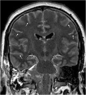
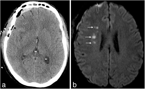
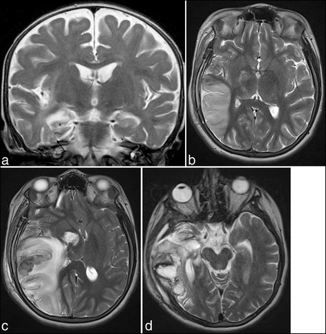
Discussion: Clinical heterogeneity represents a diagnostic challenge in the postoperative setting. Cerebral magnetic resonance imaging showed typical findings in a minority of patients only, whereas HSV-specific polymerase chain reaction on the cerebrospinal fluid proved to be a valuable test. The postoperative viral pathophysiology remains a subject of debate. The rate of adverse outcome is high and early antiviral treatment seems to be a strong predictor of clinical outcome.
Conclusion: We recommend early empirical treatment for any patient presenting with post-neurosurgical lymphocytic meningo-encephalitis, and prophylactic antiviral treatment for patients with a history of previous HSVE who will undergo a neurosurgical procedure.
Keywords: Complication; Encephalitis; Herpes simplex virus; Meningitis; Neurosurgery; Postoperative.
Figures
References
-
- Moon S, Kim T, Lee E. Comparison of clinical manifestations, outcomes and cerebrospinal fluid findings between herpes simplex type 1 and type 2 central nervous system infections in adults. J. Med. … [Internet]. 2014 [cited 2015 Jan 3];86:1766–71. Available from: http://www.ncbi.nlm.nih.gov/pubmed/25042344 - PubMed
-
- Widener RW, Whitley RJ. Herpes simplex virus. Handb. Clin. Neurol. [Internet]. 2014 [cited 2015 Jul 5];123:251–63. Available from: http://www.ncbi.nlm.nih.gov/pubmed/25015489 - PubMed
Publication types
MeSH terms
LinkOut - more resources
Full Text Sources
Other Literature Sources
Medical
