Review
. 2016 Jul;9(7):629-37.
doi: 10.1080/17474086.2016.1191944. Epub 2016 Jun 24.

Why factor XI deficiency is a clinical concern

Affiliations
Review

Why factor XI deficiency is a clinical concern

Allison P Wheeler et al. Expert Rev Hematol. 2016 Jul.

Abstract

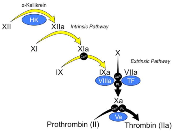
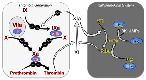
Introduction: Inherited fXI deficiency has been an enigma since its discovery in 1953. The variable and relatively mild symptoms in patients with even the most severe form of the disorder seem out of step with the marked abnormalities in standard clotting assays. Indeed, the contribution of factor XI to hemostasis in an individual is not adequately assessed by techniques available in modern clinical laboratories.

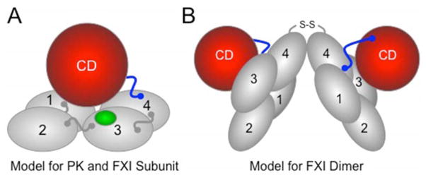
Areas covered: We discuss clinical studies, genetic/genomic analyses, and advances in laboratory medicine that are reshaping our views on the role of factor XI in pathologic coagulation. We review how the disorder associated with factor XI deficiency has contributed to changes in blood coagulation models, and discuss the complex genetics of the deficiency state and its relationship to bleeding. Finally, we cover new laboratory approaches that may distinguish deficient patients who are prone to bleeding from those without such predisposition. Expert commentary: Advances in understanding the biology of factor XI have led to modifications in treatment of factor XI-deficient patients. Factor replacement is used more judiciously, and alternative approaches are gaining favor. In the future, better laboratory tests may allow us to target therapy to those patients who would benefit most.

Keywords: Factor XI; contact activation; factor XI deficiency; factor XII; prekallikrein.


Figures

Figure 1
Figure 1. Traditional Cascade-Waterfall Model of Thrombin Generation
Black Roman numerals indicate inactive zymogens of plasma proteases, and the lowercase “a” indicates active protease. Co-factors are indicated in blue ovals. Requirements in some reactions for calcium ions (Ca2+) and phospholipid (PL) are indicated. In the activated partial thromboplastin time (aPTT assay), coagulation is initiated by activation of fXII on a charged surface in a process called contact activation, which involves the protease α-kallikrein and the cofactor high molecular weight-kininogen (HK). FXIIa triggers converting of fXI to fXIa, and fXIa in turn converts fIX to fIXa. The series of reactions indicated by yellow arrows are referred to as the intrinsic pathway. The intrinsic pathway converts fX to fXa. FXa then cleaves prothrombin to generation thrombin. The intrinsic pathway can be bypassed by the extrinsic pathway, which is comprised of fVIIa in complex with the cofactor tissue factor (TF). Activation of fX through the extrinsic pathway is the basis of the prothrombin time (PT) assay.
Figure 2
Figure 2. Current Models of Thrombin Generation and Contact Activation
Black Roman numerals indicate inactive zymogens of plasma proteases, and a lowercase “a” indicates active protease. Co-factors are indicated in blue ovals. Requirements for calcium ions (Ca2+) and phospholipid (PL) in some reactions are indicated. (Left Panel) Tissue Factor-Initiated Thrombin Generation. Thrombin generation is initiated by activation of fX by the fVIIa/TF complex. FXa then converts prothrombin to thrombin in the presence of fVa. FVIIa/TF also activates fIX, and fIXa is responsible for sustaining fX and prothrombin activation [22]. The reactions indicated by the black arrows form the core of the thrombin generation mechanism in vertebrate animals [30]. Complete absence of one of the proteins highlighted in red causes either a severe bleeding disorder or is not compatible with life. FXI is a coagulation protein found only in mammals [31]. FXI provides another mechanism for fIX activation (white arrow) that supplements fIX activation by fVIIa/TF [34]. It is thought that fXI is activated during hemostasis by thrombin in a reaction requiring an anionic cofactor [24,25]. Polyphosphate (polymerized inorganic phosphate), which is released from platelets upon activation, is a leading candidate for such a cofactor [28]. Note that fXI during hemostasis is not thought to require fXIIa. (Right Panel) Contact Activation (Kallikrein-Kinin System). Exposure of blood to a variety of artificial and biologic surfaces triggers contact activation [21,33]. FXII and prekallikrein (PK) bind to the surface and convert each other to fXIIa and α-kallikrein. High molecular weight-kininogen (HK) is a cofactor for the reaction, facilitating PK binding to the surface. FXIIa can activate fXI leading to thrombin generation. There is also evidence that fXIa, like α-kallikrein, can activate fXII [51]. Contact activation triggers thrombin generation in the aPTT assay, but it is doubtful that it contributes to hemostasis, as congenital fXII, PK or HK deficiency does not cause a bleeding disorder. Because of this, fXII, PK and HK are often considered to form a system separate from fXI referred to as the kallikrein-kinin system (KKS components highlighted in yellow) [21,33]. Activation of the KKS results in cleavage of HK by α-kallikrein generating the potent vasoactive peptide bradykinin (BK) and antimicrobial peptides (AMPs) that likely play a role in host-defense. In this figure, gray arrows indicate reactions that are enhanced by polyanions such as polyphosphate, DNA and RNA.
Figure 3
Figure 3. Models for Prekallikrein and Factor XI
(A) PK and fXI subunit structure. PK and fXI are homologs [31]. Each subunit for these proteins is comprised of four apple domains (1 through 4) and a trypsin-like catalytic domain (CD) [34,35]. The apple domains form a planar disk on which the catalytic domain rests. Activation of PK and fXI by fXIIa occurs by a single proteolytic cleavage within the portion of the proteins indicated in blue. The sequence around the fXIIa cleavage site on fXI differs from that in PK, allowing fXI to also be activated by thrombin [26]. There is a fIX binding site on the fXI apple 3 domain (indicated in green) that is not present on PK [36]. The fIX binding site is not exposed in fXI, but becomes available for fIX binding when fXI is converted to fXIa. (B) The factor XI dimer. FXI is a homodimeric protein in all species in which it has been evaluated [34,35]. The apple 4 domain serves as the interface for two fXI subunits which are oriented at a 70° angle to each other [34] In most species, a disulfide bond (S-S) connects the subunits. While dimeric and monomeric forms of fXI behave similarly in aPTT assays, the monomeric forms perform poorly in mouse thrombosis models, indicating that the dimeric conformation serves a critical function in vivo [29].

References

    1. Rosenthal RL, Dreskin OH, Rosenthal N. New hemophilia-like disease caused by deficiency of a third plasma thromboplastin factor. Proc Soc Exp Biol Med. 1953;82:171–4. - PubMed
    1. Rosenthal RL, Dreskin OH, Rosenthal N. Plasma thromboplastin antecedent (PTA) deficiency; clinical, coagulation, therapeutic and hereditary aspects of a new hemophilia-like disease. Blood. 1955;10:120–31. - PubMed
    1. Wright IS. The nomenclature of blood clotting factors. Canad Med Ass J. 1962;86:373–4. - PMC - PubMed
    1. Duga S, Salomon O. Congenital factor XI deficiency: an update. Sem Thromb Hemost. 2013;39:621–31. - PubMed
    1. Santoro C, Di Mauro R, Baldacci E, et al. Bleeding phenotype and correlation with factor XI (FXI) activity in congenital FXI deficiency: results of a retrospective study from a single centre. Haemophilia. 2015;21:496–501. - PubMed

LinkOut - more resources