. 2016 Jul 17;428(14):2814-31.
doi: 10.1016/j.jmb.2016.05.013. Epub 2016 May 21.

The Structure-Specific Recognition Protein 1 Associates with Lens Epithelium-Derived Growth Factor Proteins and Modulates HIV-1 Replication

Affiliations

The Structure-Specific Recognition Protein 1 Associates with Lens Epithelium-Derived Growth Factor Proteins and Modulates HIV-1 Replication

Angelica P Lopez et al. J Mol Biol. .

Abstract

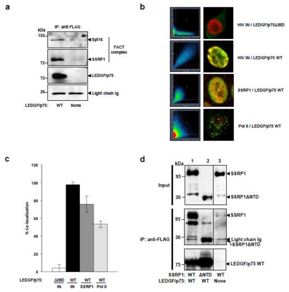
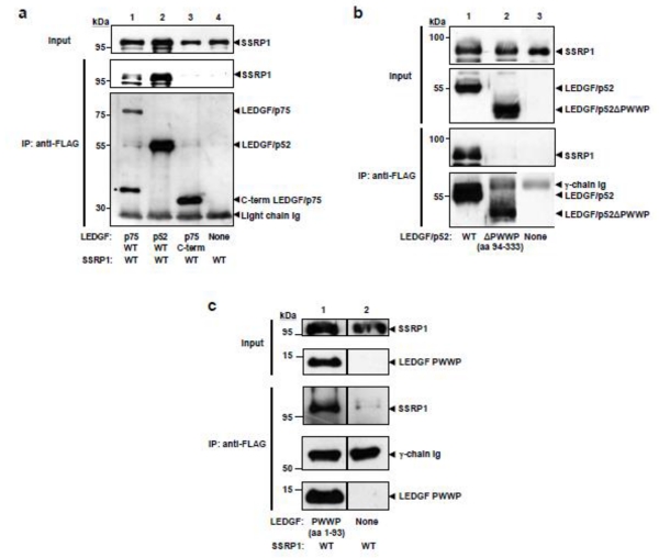
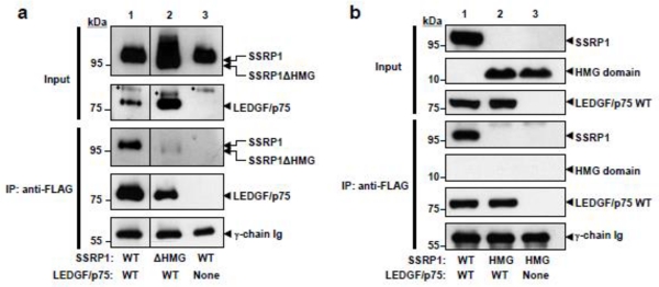
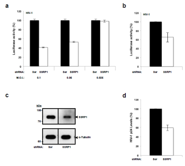
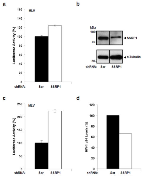
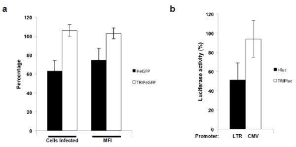
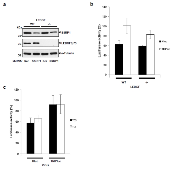
The lens epithelium-derived growth factor p75 (LEDGF/p75) is a chromatin-bound protein essential for efficient lentiviral integration. Genome-wide studies have located LEDGF/p75 inside actively transcribed genes where it mediates lentiviral integration. Although its role in HIV-1 integration is clearly established, the role of LEDGF/p75-associated proteins in HIV-1 infection remains unexplored. Using protein-protein interaction assays, we demonstrated that LEDGF/p75 complexes with a chromatin-remodeling complex facilitates chromatin transcription (FACT), a heterodimer of the structure-specific recognition protein 1 (SSRP1) and the human homolog of suppressor of Ty 16 (hSpt16). Detailed analysis of the interaction of LEDGF/p75 with the FACT complex indicates that LEDGF/p75 interacts with SSRP1 in an hSpt16-independent manner that requires the PWWP domain of LEDGF proteins and the HMG domain of SSRP1. Functional characterizations demonstrate a LEDGF/p75-independent role of SSRP1 in the regulation of HIV-1 replication. shRNA-mediated partial knockdown of SSRP1 reduces HIV-1 infection, but not Murine Leukemia Virus, in human CD4(+) T cells. Similarly, SSRP1 knockdown affects infection by HIV-1-derived viruses that express genes from the viral LTR but not from an internal immediate-early CMV promoter, suggesting a role of SSRP1 in LTR-driven gene expression but not in viral DNA integration. Together, our data demonstrate for the first time the association of LEDGF proteins with the FACT complex and give further support to a role of SSRP1 in HIV-1 infection.

Keywords: FACT complex; HIV-1 cofactor; HIV-1 replication; HMG domain; PWWP domain.


Figures

Figure 1
Figure 1. Interaction of LEDGF/p75 with the FACT complex
(a) Chromatin-bound proteins were isolated from TL3 and TL3 LEDGF/p75 WT cells by DNase treatment and FLAG-tagged LEDGF/p75 was immunoprecipitated from this subcellular fraction with an anti-FLAG mAb antibody. The presence of the FACT complex (hSpt16 and SSRP1) was evaluated in the immunoprecipitated proteins by immunoblotting with specific antibodies. (b) Quantitative confocal microscopy co-localization of LEDGF/p75 with SSRP1. Panels 1 and 2 are controls and represent the co-localization of HIV integrase with LEDGF/p75 mutant lacking the integrase-binding domain (ΔIBD) or LEDGF/p75 wild type (WT). LEDGF/p75-deficient HEK293T cells stably expressing eGFP-tagged HIV-1 integrase were transiently transfected with LEDGF/p75ΔIBD or WT. LEDGF/p75 proteins were detected with an anti-LEDGF antibody. The lower panels represent the co-localization of endogenous LEDGF/p75 with endogenous SSRP1 or Pol II in HeLa cells after immunostaining with specific antibodies. (c) Quantification of co-localization data in (b). Standard deviations indicated co-localization values found in ten different cells randomly selected from a field representative of ten different random areas of the microscope slide. (d) HEK293T cells were co-transfected with plasmids expressing: FLAG-tagged LEDGF/p75 and Myc-tagged SSRP1 WT (lane 1), FLAG-tagged LEDGF/p75 and Myc-tagged SSRP1ΔNTD (lane 2), or Myc-tagged SSRP1 WT and an empty plasmid (lane 3). Samples were analyzed in the same gel, the line dividing lanes 2 and 3 indicate that lanes in between were removed for comparison in this figure. In 1d, (*) indicates degradation products of SSRP1
Figure 2
Figure 2. Mapping the LEDGF regions implicated in SSRP1 binding
(a) si1340/1428 cells were co-transfected with plasmids encoding Myc-SSRP1 and either LEDGF/p75-FLAG (lane 1), LEDGF/p52-FLAG (lane 2), C-terminal LEDGF/p75-FLAG (lane 3), or an empty expression plasmid (lane 4). Immunoprecipitation analyses were performed as previously described in figure legend 1a. (*) Marks degradation products of LEDGF/p52-FLAG. (b) Implication of PWWP domain in the interaction of LEDGF/p52 with SSRP1. Cell lysates were obtained from si1340/1428 cells co-transfected with plasmids expressing Myc-SSRP1 and either LEDGF/p52-FLAG (lane 1), LEDGF/p52ΔPWWP-FLAG (lane 2), or an empty plasmid (lane 3). Immunoprecipitations were performed as previously described. (c) si1340/1428 cells were co-transfected with plasmids expressing Myc-SSRP1 and PWWP-FLAG (lane 1) or an empty plasmid (lane 2). Immunoprecipitations were performed as previously described in figure legend 1a. Results in (c) are representative of two independent experiments. The line separating lanes 1 and 2 indicate that lanes of the gel in between contained irrelevant samples that were removed.
Figure 3
Figure 3. Immunoprecipitation analyses of the interaction of SSRP1 mutants with LEDGF/p75
(a) HEK293T cells were co-transfected with plasmids expressing: FLAG-tagged LEDGF/p75 and Myc-tagged SSRP1 WT (lane 1), FLAG-tagged LEDGF/p75 and Myc-tagged SSRP1ΔHMG (lane 2), or Myc-tagged SSRP1 WT and an empty plasmid (lane 3). Results in (a) are representative of three independent experiments. The vertical lines separating lanes 1 and 2 indicate that samples were in the same gel but not in adjacent positions. (*) Denotes residual SSRP1 resulting from partial antibody stripping. (b) HEK293T cells were co-transfected with plasmids encoding LEDGF/p75-FLAG and SSRP1 WT-Myc (lane 1) or HMG-Myc (lane 2). Lane 3 was co-transfected with SSRP1 WT-Myc and an empty plasmid. S1 and S2 fractions were obtained from the transfected cells and mixed. Mixed fractions were immunoprecipitated as described in figure legend 1a. Results are representative of two independent experiments.
Figure 4
Figure 4. Effect of SSRP1 partial knockdown on HIV-1 infection
(a and b) SupT1 cells were transduced at different MOIs with a lentiviral vector expressing a Scrambled or SSRP1-specific shRNA and three days later were infected with single-round HIV-1 expressing luciferase. Three days post-infection luciferase and ATP levels were measured. Luciferase readings in control cells infected with Hluc in (a) and (b) were around 5×10³ relative light untis (RLU)/ml and background readings were approximately 0.02 RLU/ml. Luciferase was normalized to ATP content in the same samples. Standard deviations in (a) represent the variability in luciferase readings of single experiments and in (b) the variability of three independent experiments at MOI 0.1. (c) The levels of SSRP1 in one of the experiments represented in (b) were determined by immunoblot analysis. α-tubulin was measured as a loading control. The level of knockdown achieved is representative of the other two infection experiments. The vertical line separating the lanes indicates that the samples were in the same electrophoresis gel and immunoblot membrane but not in adjacent positions. (d) SupT1 cells were transduced with either the scrambled or SSRP1-specific shRNA expressing lentiviral virus and three days later were infected with HIV-1NL4-3. After twenty-four hours, infected cells were extensively washed to remove input HIV-1NL4-3 virus and 96 hours post-infection the supernatant was harvested and HIV-1 p24 quantified by ELISA. Standard deviations represent data from three different experiments.
Figure 5
Figure 5. Influence of partial knockdown of SSRP1 on MLV and HIV-1 infections
(a) SupT1 cells were transduced with a lentiviral vector encoding a Scrambled or SSRP1-specific shRNA, three days later the cells were infected with an MLV-derived virus expressing luciferase, and seventy-two hours post-infection, luciferase was measured. (b-d) SupT1 cells were transduced at MOI 1 with a lentiviral vector encoding a Scrambled or SSRP1-specific shRNA and three days later one-third of the cells were analyzed for SSRP1 expression by immunoblot (b) and the remaining cells were infected with MLV (c) or HIV-1NL4-3 (d). Three days after infection, luciferase activity was measured in the MLV-infected cells (c) and HIV-1 replication was determined by quantifying HIV-1 p24 in the supernatant of the HIV-1NL4-3 infected cells by ELISA (d). Standard deviation in (a) and (c) represent the variability of luciferase activity readings from single experiments. The vertical line separating the lanes in (b) indicates that the samples, although in the same electrophoresis gel and immunoblot membrane, were not in adjacent positions. Luciferase readings in control cells infected with MLVluc in (a) and (c) were around 8 RLU/ml and background readings were approximately 0.02 RLU/ml.
Figure 6
Figure 6. Influence of partial knockdown of SSRP1 on HIV-1 infection carrying different promoters
(a and b) SupT1 cells were transduced with a lentiviral vector encoding a Scrambled or SSRP1-specific shRNA, three days later the cells were infected with HIV-derived viruses expressing eGFP or luciferase from the HIV-1 LTR (HeGFP/Hluc) or from an internal immediate early CMV promoter (TRIPeGFP/TRIPluc). Seventy-two hours post-infection, luciferase or eGFP were measured. Luciferase activity measured in the cells transduced with control shRNAs was considered 100% values. Standard deviations represent the variability of four independent experiments.
Figure 7
Figure 7. Consequence of SSRP1 partial knockdown in LEDGF/p75-deficient cells on HIV-1 infections
(a and b) LEDGF/p75 wild type or knockout Nalm-6 cells were transduced with a lentiviral vector expressing a Scrambled or SSRP1-specific shRNA and after three days one-third of these cells were analyzed for SSRP1 and LEDGF/p75 expression by immunoblot in (a). The remaining cells were infected with HIV-1-derived viruses expressing luciferase from the HIV-1 LTR (Hluc) or an internal immediate CMV promoter (TRIPluc). Seventy-two hours post-infection, luciferase and viability were measured in (b). (c) LEDGF/p75-deficent (TL3) and control (TC3) cells were transduced with a lentiviral vector encoding a Scrambled or SSRP1-specific shRNA, three days later the cells were infected with Hluc or TRIPluc and seventy-two hours post-infection, luciferase was measured. Luciferase activity measured in the cells transduced with control shRNAs was considered 100% values. Standard deviations represent the variability of six (b) or four (c) independent transduction/infection experiments.

Similar articles

Cited by

References

    1. Ge H, Si Y, Roeder RG. Isolation of cDNAs encoding novel transcription coactivators p52 and p75 reveals an alternate regulatory mechanism of transcriptional activation. Embo J. 1998;17:6723–9. - PMC - PubMed
    1. Ge H, Si Y, Wolffe AP. A novel transcriptional coactivator, p52, functionally interacts with the essential splicing factor ASF/SF2. Mol Cell. 1998;2:751–9. - PubMed
    1. Kubo E, Singh DP, Fatma N, Shinohara T, Zelenka P, Reddy VN, Chylack LT. Cellular distribution of lens epithelium-derived growth factor (LEDGF) in the rat eye: loss of LEDGF from nuclei of differentiating cells. Histochem Cell Biol. 2003;119:289–99. - PubMed
    1. Llano M, Morrison J, Poeschla EM. Virological and cellular roles of the transcriptional coactivator LEDGF/p75. Curr Top Microbiol Immunol. 2009;339:125–46. - PMC - PubMed
    1. Llano M, Vanegas M, Hutchins N, Thompson D, Delgado S, Poeschla EM. Identification and characterization of the chromatin-binding domains of the HIV-1 integrase interactor LEDGF/p75. J Mol Biol. 2006;360:760–73. - PubMed

MeSH terms

Substances

LinkOut - more resources