Review
. 2016;16(3):156-67.
doi: 10.2174/1566523216666160524144041.

Tet-On Systems For Doxycycline-inducible Gene Expression

Affiliations
Review

Tet-On Systems For Doxycycline-inducible Gene Expression

Atze T Das et al. Curr Gene Ther. 2016.

Abstract

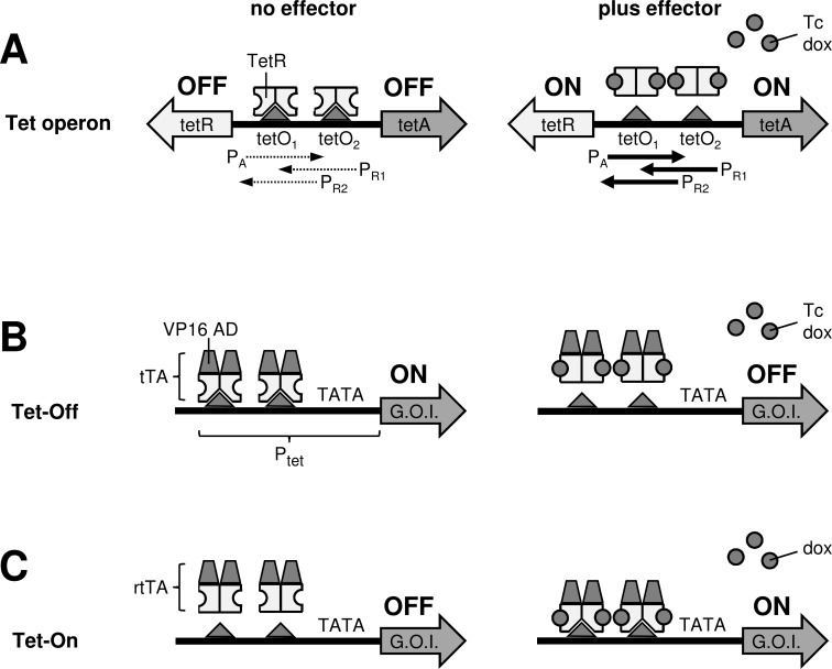
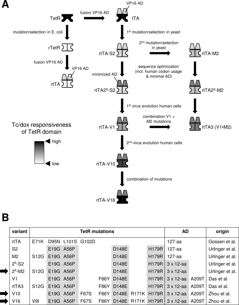
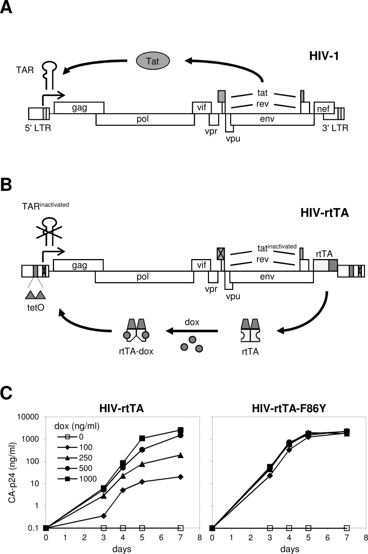
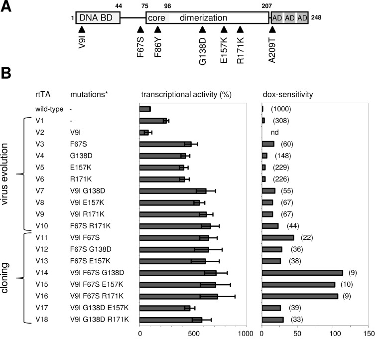
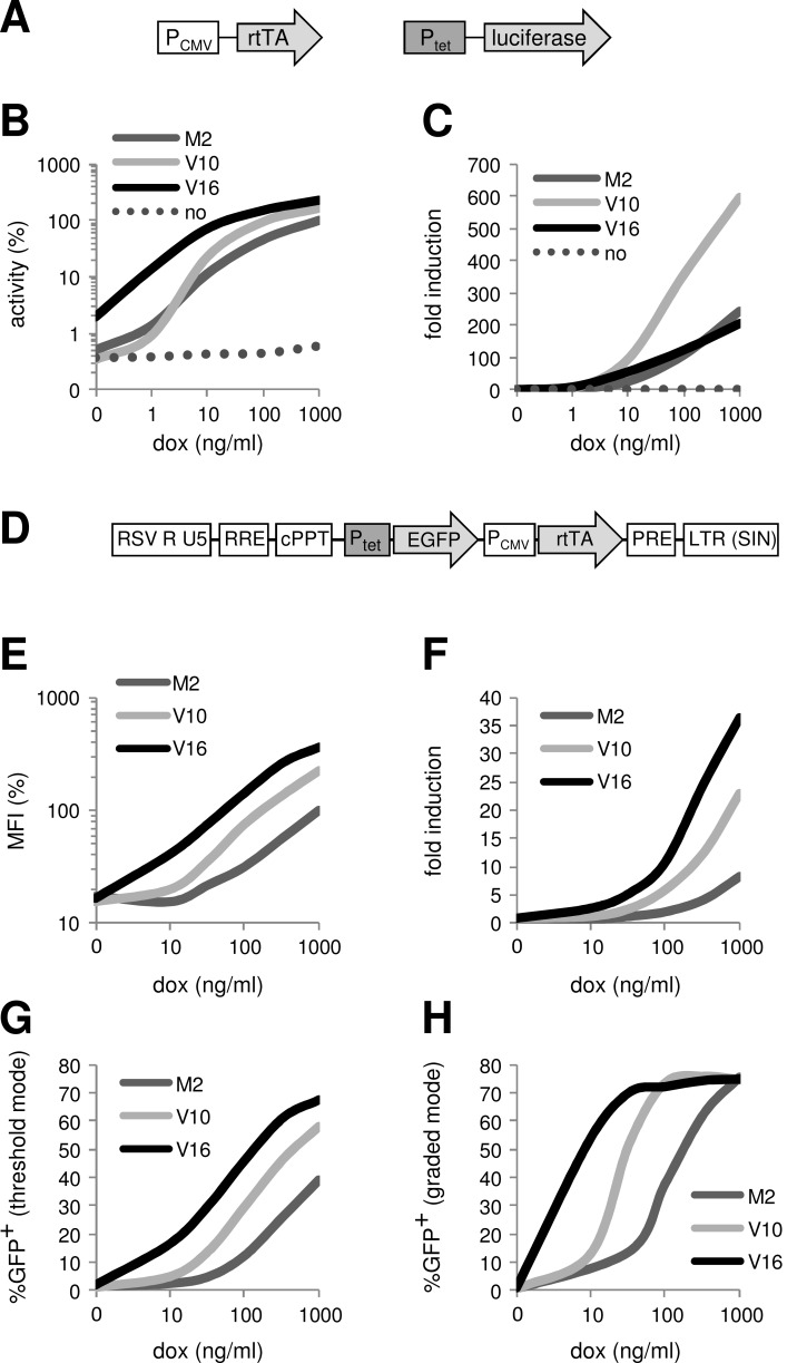
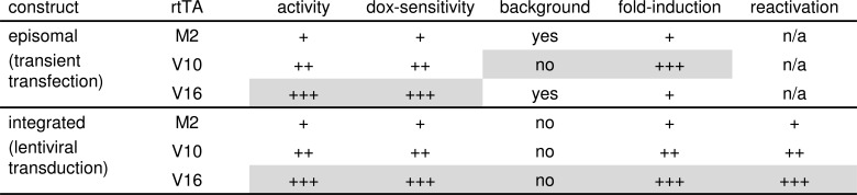
The tetracycline-controlled Tet-Off and Tet-On gene expression systems are used to regulate the activity of genes in eukaryotic cells in diverse settings, varying from basic biological research to biotechnology and gene therapy applications. These systems are based on regulatory elements that control the activity of the tetracycline-resistance operon in bacteria. The Tet-Off system allows silencing of gene expression by administration of tetracycline (Tc) or tetracycline-derivatives like doxycycline (dox), whereas the Tet-On system allows activation of gene expression by dox. Since the initial design and construction of the original Tet-system, these bacterium-derived systems have been significantly improved for their function in eukaryotic cells. We here review how a dox-controlled HIV-1 variant was designed and used to greatly improve the activity and dox-sensitivity of the rtTA transcriptional activator component of the Tet-On system. These optimized rtTA variants require less dox for activation, which will reduce side effects and allow gene control in tissues where a relatively low dox level can be reached, such as the brain.


Figures

Fig. (1)
Fig. (1)
Tc-controlled regulation of gene expression. (A) Tn10 tet operon. In E. coli, TetR binds as a dimer to the tetO1 and tetO2 sites in the Tn10 tet operon. This interaction blocks the activity of the underlying promoters (PA, PR1 and PR2) and inhibits transcription of the tetA and tetR genes. Binding of Tc or dox triggers a conformational switch in TetR that prevents tetO binding and results in the activation of TetA and TetR production. (B) The Tet-Off system. Fusion of TetR to the activation domain of the herpes simplex virus VP16 protein (VP16 AD) resulted in the Tc-controlled transcriptional activator (tTA). Binding of tTA to the Ptet promoter that consists of 7 tetO sequences fused to a minimal TATA-box containing eukaryotic promoter, activates expression of the downstream positioned gene-of-interest (G.O.I.) Binding of Tc or dox induces a conformational change in the TetR domain of tTA, which prevents tetO binding and switches gene expression off. (C) The Tet-On system. The reverse-tTA (rtTA) variant exhibits a reverse phenotype and does not bind tetO in the absence of an effector. Binding of dox triggers a conformational switch in rtTA, which allows tetO binding. Subsequent activation of the Ptet promoter drives expression of the downstream positioned gene. The initial version of rtTA had a low affinity for Tc and was not activated by this compound.
Fig. (2)
Fig. (2)
Construction and optimization of the Tet-On system. (A) Schematic overview of the development of rtTA variants by rational design, random mutation and screening in E. coli and yeast (S. cerevisiae) and virus evolution in human cells. See text for details. The color of the TetR domain reflects the effector responsiveness (white, low Tc/dox response; black, high Tc/dox response). (B) Mutations in the TetR moiety and activation domain (AD) in different rtTA variants. The activity of the variants indicated with an arrow was directly compared in transiently transfected and stably transduced cells (Figs. 5, 6).
Fig. (3)
Fig. (3)
The dox-dependent HIV-rtTA virus. (A) Transcription of wild-type HIV-1 is activated by the binding of the viral Tat protein to the TAR hairpin structure that is present at the 5’ end of nascent RNA transcripts. This Tat-TAR axis thus controls viral gene expression and replication. (B) In HIV-rtTA, Tat and TAR are inactivated by mutation and functionally replaced by the rtTA-tetO components of the Tet-On system, by insertion of the rtTA gene at the site of the nef gene and insertion of tetO sequences in the LTR-promoter region. Transcription of this HIV-1 variant is activated by the binding of rtTA to the tetO-LTR promoter. Because rtTA binds the tetO sites exclusively in the presence of dox, HIV-rtTA does not replicate in the absence of this effector. (C) Dox-controlled replication of HIV-rtTA strains with the original rtTA-2S-S2 gene (left panel) and the F86Y-mutated variant (right panel) in SupT1 T-cells at different dox-concentrations. Virus replication is monitored by measuring the viral capsid protein (CA-p24) in the culture supernatant. Reproduced from [67] (© 2004, the American Society for Biochemistry and Molecular Biology).
Fig. (4)
Fig. (4)
Optimization of the Tet-On system through virus evolution. (A) Amino acid substitutions observed in rtTA-2S-S2 (wild-type) upon long-term culturing of HIV-rtTA in human SupT1 T-cells. (B) The transcriptional activity (at 1000 ng/ml dox) and dox-sensitivity of the wild-type, naturally evolved (V1-V10) and constructed (V11-V18) rtTA variants was measured in HeLa X1/6 cells that contain chromosomally integrated copies of the Ptet-luciferase reporter construct. The wild-type rtTA activity at 1000 ng/ml dox was set at 100%. The activity measured at different dox concentrations was used to calculate the dox concentration that each rtTA variant needs to reach an activity comparable to that of the wild-type rtTA at 1000 ng/ml dox. These concentrations are indicated between brackets in the right panel (nd, not determined), and were used to calculate the dox-sensitivity for each rtTA variant (dox-sensitivity of wild-type rtTA set at 1). *, V1-V18 carry the F86Y and A209T mutations in addition to the shown mutations. Reproduced from [69] (© 2006, Zhou et al.).
Fig. (5)
Fig. (5)
Comparison of rtTA variants in transiently transfected and stably transduced cells. (A-C) Different human cell lines (HeLa cervix carcinoma and HEK293 embryonal kidney cells) were transfected with low and high amounts of CMV enhancer/promoter-driven PCMV-rtTA (either rtTA2S-M2 [M2], rtTA-V10 or rtTA-V16) and dox-rtTA activated Ptet-luciferase constructs (constructs shown in A), and cultured in the presence of 0 to 1000 ng/ml dox. (B) The luciferase level measured 2 days after transfection reflects the rtTA activity. The activity of M2 at 1000 ng/ml dox was set at 100%. The average of the values obtained for different experimental conditions (different cells and different amounts of DNA) [70] is shown to illustrate the differences between the rtTA variants (no, transfection of cells with the empty vector instead of the rtTA plasmid). The original data for each cell line and amount of DNA are presented in [70]. (C) Fold induction levels, calculated as the ratio between the rtTA activity at the indicated dox concentration and the activity in the absence of dox (0 ng/ml). (D-H) Different human cell lines (SupT1, HeLa, HepG2 hepatocellular carcinoma and SJNB-8 neuroblastoma cells) were transduced with a lentiviral vector containing Ptet-d2EGFP and PCMV-rtTA (M2, V10 or V16) cassettes (construct shown in D). Cells were cultured with dox for 3 days. GFP-positive (GFP+) cells were sorted and cultured without dox for 6 days to switch off GFP expression. The isolated transduced cells were subsequently cultured with different dox concentrations for 3 days, after which the intracellular GFP level was analyzed. (E) The mean fluorescent intensity (MFI) of the total cell population reflects the rtTA activity. The activity of M2 at 1000 ng/ml dox was set at 100%. The average of the values obtained with the different cells (original data published in [70]) is shown to illustrate the differences between the rtTA variants. (F) Fold induction levels, calculated as the ratio between the MFI at the indicated dox concentration and the MFI in the absence of dox (0 ng/ml). (G) The average percentage of GFP+ cells measured for the SupT1, HeLa and SJNB-8 cells that express GFP predominantly in a threshold mode (i.e. increasing the dox concentration activated GFP expression in more cells, rather than that it increased the fluorescence intensity of the GFP+ cells [89]; original data published in [70]). (H) The percentage of GFP+ cells measured for HepG2 cells that express GFP predominantly in a graded mode (i.e. increasing the dox concentration predominantly increased the fluorescence intensity of the GFP+ cells [89]).
Fig. (6)
Fig. (6)
Identification of the optimal Tet-On system for different applications. Direct comparison of the activity of the rtTA-2S-M2 (M2), rtTA-V10 and rtTA-V16 variants in transiently transfected and stably transduced cells (as shown in Fig. 5) revealed their quality with respect to different parameters. +, good; ++, better; +++, best; n/a, not applicable.

References

    1. Fussenegger M. The impact of mammalian gene regulation concepts on functional genomic research, metabolic engineering, and advanced gene therapies. Biotechnol. Prog. 2001;17(1):1–51. doi: 10.1021/bp000129c. - DOI - PubMed
    1. Gossen M., Bujard H. Tetracyclines in the control of gene expression in eukaryotes. In: Nelson M., Hillen W., Greenwald R.A., editors. Tetracyclines in biology, chemistry and medicine. Basel: Birkhäuser Verlag; 2001. pp. 139–157. - DOI
    1. Baron U., Bujard H. Tet repressor-based system for regulated gene expression in eukaryotic cells: principles and advances. Methods Enzymol. 2000;327:401–421. doi: 10.1016/S0076-6879(00)27292-3. - DOI - PubMed
    1. Berens C., Hillen W. Gene regulation by tetracyclines. Constraints of resistance regulation in bacteria shape TetR for application in eukaryotes. Eur. J. Biochem. 2003;270(15):3109–3121. doi: 10.1046/j.1432-1033.2003.03694.x. - DOI - PubMed
    1. Roberts M.C. Update on acquired tetracycline resistance genes. FEMS Microbiol. Lett. 2005;245(2):195–203. doi: 10.1016/j.femsle.2005.02.034. - DOI - PubMed