. 2016 May 24:6:26571.
doi: 10.1038/srep26571.

Cold-inducible RNA-binding protein causes endothelial dysfunction via activation of Nlrp3 inflammasome

Affiliations

Cold-inducible RNA-binding protein causes endothelial dysfunction via activation of Nlrp3 inflammasome

Weng-Lang Yang et al. Sci Rep. .

Abstract

Cold-inducible RNA-binding protein (CIRP) is a damage-associated molecular pattern (DAMP) molecule which stimulates proinflammatory cytokine release in hemorrhage and sepsis. Under these medical conditions, disruption of endothelial homeostasis and barrier integrity, typically induced by proinflammatory cytokines, is an important factor contributing to morbidity and mortality. However, the role of CIRP in causing endothelial dysfunction has not been investigated. In this study, we show that intravenous injection of recombinant murine CIRP (rmCIRP) in C57BL/6 mice causes lung injury, evidenced by vascular leakage, edema, increased leukocyte infiltration and cytokine production in the lung tissue. The CIRP-induced lung damage is accompanied with endothelial cell (EC) activation marked by upregulation of cell-surface adhesion molecules E-selectin and ICAM-1. Using in vitro primary mouse lung vascular ECs (MLVECs), we demonstrate that rmCIRP treatment directly increases the ICAM-1 protein expression and activates NAD(P)H oxidase in MLVECs. Importantly, CIRP stimulates the assembly and activation of Nlrp3 inflammasome in MLVECs accompanied with caspase-1 activation, IL-1β release and induction of proinflammatory cell death pyroptosis. Finally, our study demonstrates CIRP-induced EC pyroptosis in the lungs of C57BL/6 mice for the first time. Taken together, the released CIRP in shock can directly activate ECs and induce EC pyroptosis to cause lung injury.


Figures

Figure 1
Figure 1. Extracellular CIRP induces vascular leakage and causes lung injury.
The C57BL/6 mice were intravenously injected with vehicle (normal saline) or recombinant murine CIRP (rmCIRP, 5 mg/kg body weight) and lungs were collected for various analysis 5 h later. (A) Representative images of the lungs stained with Evans blue dye (EBD) post-vehicle or rmCIRP injections. (B) Colorimetric analysis of EBD extracted from stained lungs. Bars represent mean ± SEM (n = 5 per group). *P < 0.05; Student’s t-test. Representative images of H&E stained lung sections from (C) vehicle or (D) rmCIRP injected mice at original magnification ×400. The qPCR analysis of (E) cytokines and (F) endothelial cell adhesion molecules in the lungs of vehicle or rmCIRP injected mice. Bars represent mean ± SEM (n = 3 per group). *P < 0.05; Student’s t-test.
Figure 2
Figure 2. CIRP activates vascular endothelial cells.
(A) Western blot analysis of ICAM-1 in the mouse lung vascular endothelial cells (MLVECs) treated with an indicated concentration of rmCIRP for 4 h. The image shown is representative of three independent experiments. Bars represent mean ± SEM (n = 3) from densitometric analysis of blots. *P < 0.05 compared to no rmCIRP; one-way ANOVA, Student-Newman-Keuls test. (B) Western blot analysis of ICAM-1 in MLVECs treated with 200 ng/ml rmCIRP for upto 8 h as indicated. The image is representative of three independent experiments. Bars represent mean ± SEM (n = 3). *P < 0.05 compared to time 0; one-way ANOVA, Student-Newman-Keuls test.
Figure 3
Figure 3. CIRP induces NAD(P)H oxidase activation.
Western blot analysis of gp91phox, phosphorylated p47phox (phospho-p47phox) and total p47phox from mouse lung vascular endothelial cells (MLVECs) immunoprecipitated with anti-p47phox antibody after treatment with (A) an indicated concentration of rmCIRP for 4 h or (B) 200 ng/ml rmCIRP for upto 8 h as indicated. The images are representative of three independent experiments.
Figure 4
Figure 4. CIRP induces Nlrp3 inflammasome activation and pyroptosis in vitro.
(A,B) Mouse lung vascular endothelial cells (MLVECs) were treated with an indicated concentration of rmCIRP for an indicated period of time. (A) The cells from the treatments were subjected to immunoprecipitation with anti-ASC antibody. Immunoblot analysis of Nlrp3, ASC and cleaved caspase-1 was performed. The images are representative of three independent experiments. (B) IL-1β levels in the cell culture media from above treatments, measured by ELISA. Bars represent mean ± SEM (n = 3). *P < 0.05 compared to no rmCIRP or 0 h; #P < 0.05 compared to 200 ng/ml rmCIRP or 1 h; one-way ANOVA, Student-Newman-Keuls test. (C,D) MLVECs were treated with no rmCIRP or 200 ng/ml rmCIRP for upto 24 h as indicated. Cells were then stained with TMR-red-Cell Death reagent and Alexa Fluor 488-FLICA-activated caspase-1 fluorescent reagent (AF488-caspase-1), followed by flow cytometry. (C) Representative dot blots showing percentages of double-stained pyroptotic MLVECs as indicated by numbers in the top right quadrant. (D) Graph showing the percentages of pyroptotic cells from flow cytometric analysis. Bars represent mean ± SEM (n = 3). *P < 0.05 compared to no rmCIRP; one-way ANOVA, Student-Newman-Keuls test.
Figure 5
Figure 5. CIRP induces endothelial cell pyroptosis in vivo.
C57BL/6 mice were intravenously injected with vehicle (normal saline) or rmCIRP (5 mg/kg body weight). After 24 h, lung tissues were harvested and subjected to fluorescent immunohistochemistry analysis. Representative images of lung tissues viewed under confocal microscopy (original magnification x600) after staining with Alexa Fluor 488- FLICA-activated caspase-1 (green), TUNEL (red), APC-E-selectin-1 (white) and Hoechst (blue). Images are representative of three independent experiments.
Figure 6
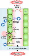
Figure 6. Model of CIRP-induced vascular endothelial cell dysfunction.
CIRP released during hemorrhagic or septic shock activates endothelial cells (EC) by upregulating adhesion molecules on them to increase infiltration of polymorphonuclear leukocytes (PMN) and produce proinflammatory cytokines and reactive oxygen species (ROS). CIRP also induces activation of Nlrp3 inflammasome and caspase-1-mediated EC pyroptosis. This series of events serve as a mechanism underlying the CIRP-induced EC dysfunction which then leads to organ injury.

References

    1. Schouten M., Wiersinga W. J., Levi M. & van der Poll T. Inflammation, endothelium, and coagulation in sepsis. J Leukoc Biol. 83, 536–545 (2008). - PubMed
    1. Diebel L. N., Liberati D. M., Ledgerwood A. M. & Lucas C. E. Changes in lymph proteome induced by hemorrhagic shock: the appearance of damage-associated molecular patterns. J Trauma Acute Care Surg. 73, 41–51 (2012). - PubMed
    1. Medzhitov R. Origin and physiological roles of inflammation. Nature. 454, 428–435 (2008). - PubMed
    1. Kaczorowski D. J., Mollen K. P., Edmonds R. & Billiar T. R. Early events in the recognition of danger signals after tissue injury. J Leukoc Biol. 83, 546–552 (2008). - PubMed
    1. Hutchins N. A., Wang F., Wang Y., Chung C. S. & Ayala A. Kupffer cells potentiate liver sinusoidal endothelial cell injury in sepsis by ligating programmed cell death ligand-1. J Leukoc Biol. 94, 963–970 (2013). - PMC - PubMed

Publication types

MeSH terms

LinkOut - more resources